





Expo
view channel
view channel
view channel
view channel
view channel
view channel
view channel
view channel
AICritical CareSurgical TechniquesPatient Care
Health ITPoint of CareBusiness
Events
Webinars

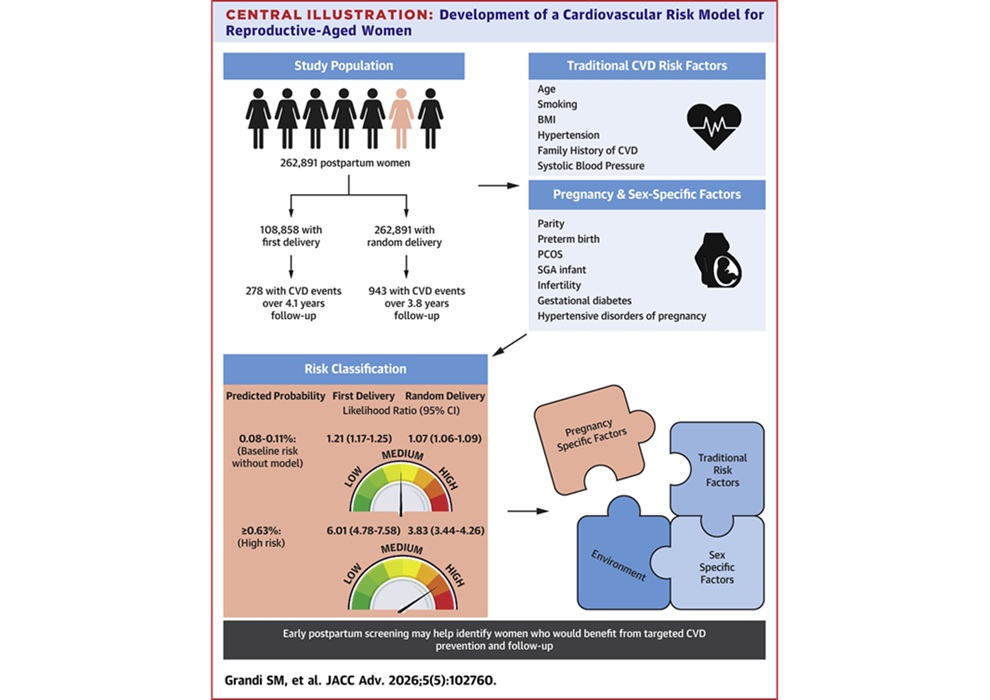
- New Risk Calculator Identifies Heart Disease Earlier in Postpartum Women
- Radiation-Free Cardiac Catheterization Improves Care for Children
- New Soft Optical Sensor Advances Heart and Brain Activity Mapping
- AI System Stabilizes Oxygen Levels More Reliably Than Standard Care
- Simple Triage Protocol Reduces Emergency Department Length of Stay
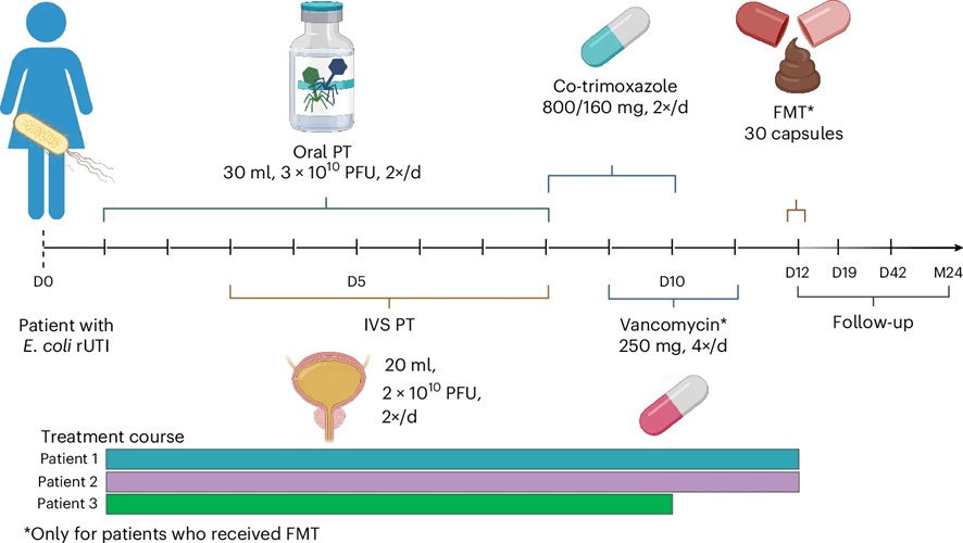
- Combined Phage and Microbiota Therapy May Reduce Recurrent Urinary Tract Infections
- New AR Technology Provides Real-Time Guidance During ACL Surgery
- New Technique Rapidly Produces Patient-Specific Vascular Grafts During Surgery
- AI Analyzes Surgical Movements to Improve Prostatectomy Outcomes
- Photostable NIR Dye Sustains Intraoperative Imaging in Cancer Surgery
- Wearable Sleep Data Predict Adherence to Pulmonary Rehabilitation
- Revolutionary Automatic IV-Line Flushing Device to Enhance Infusion Care
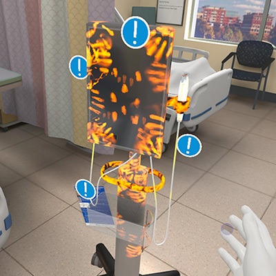
- VR Training Tool Combats Contamination of Portable Medical Equipment
- Portable Biosensor Platform to Reduce Hospital-Acquired Infections
- First-Of-Its-Kind Portable Germicidal Light Technology Disinfects High-Touch Clinical Surfaces in Seconds
- AI Accelerates Individualized Dosimetry for Prostate Cancer Treatment
- Automated Breast Ultrasound Platform Improves Dense Breast Screening
- ‘Local Brain Age’ AI Mapping Tool Reveals Patterns of Cognitive Impairment
- Rapid AI System Detects and Classifies Brain Tumor Subtypes on MRI
- PET/CT Improves Diagnosis and Management of Fever of Unknown Origin
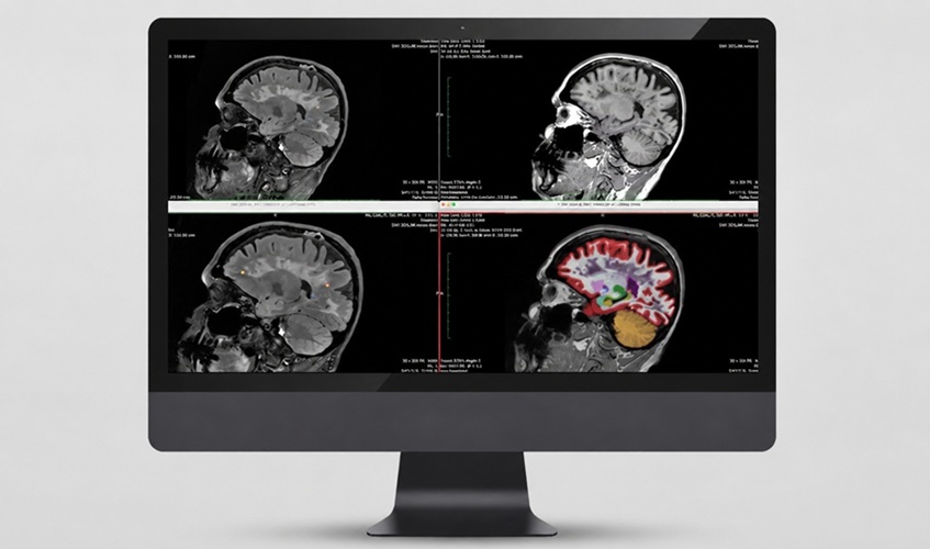
- AI Care Platform Adds Quantitative MRI Analysis for Multiple Sclerosis
- Danaher Completes Acquisition of Masimo to Expand Patient Monitoring Capabilities
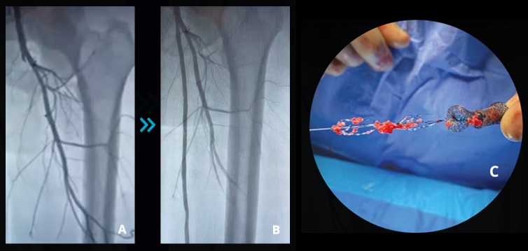
- Endologix Adds FDA-Cleared Peripheral Thrombectomy System
- Artivion Adds FDA-Approved NEXUS System to Aortic Arch Portfolio
- Olympus Partnership Aims to Expand Access to Robot-Assisted Endoscopic Therapy
- New Cloud Platform Streamlines Capsule Endoscopy Workflow and Reporting
- Smartwatch Monitoring Identifies Abnormal Vital Signs Before Routine Checks
- Weekly Remote Symptom Monitoring Improves Symptom Control in Advanced Cancer
- Digital Heart Model Supports Targeted Ablation in Atrial Fibrillation
- AI Framework Helps Clinicians Create Trustworthy Risk Prediction Tools
- AI-Powered Brain-Body Interface Supports Movement and Sensation Recovery
- New AI Tool Identifies Support Needs in Spinal Cord Stimulation
- AI Tool Predicts Chronic Kidney Disease Risk in Diabetes
- AI Trends Report Guides Responsible, Effective Healthcare Deployment
- Privacy-Preserving AI Protects Sensitive Information in ECG Data
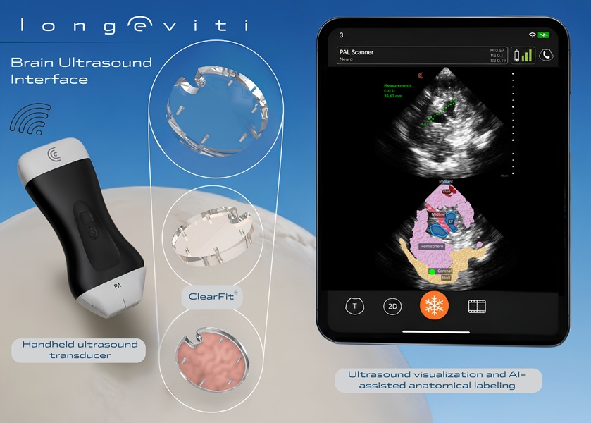
- New Brain Ultrasound Platform Enables Bedside Postoperative Imaging
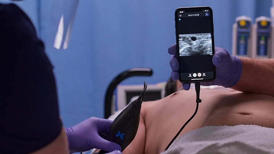
- Handheld Ultrasound Expands Point-of-Care Imaging Access in Brazil
- AI Dermatology Platform Targets Early Detection of Non-Melanoma Skin Cancer
- Handheld AI Device for Point-of-Care Skin Lesion Assessment Receives CE Mark
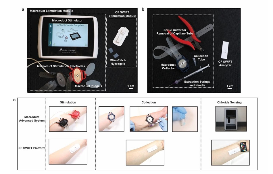
- Portable Immunoassay System Advances Toward Point-of-Care Biomarker Testing
 Expo
Expo
- New Risk Calculator Identifies Heart Disease Earlier in Postpartum Women
- Radiation-Free Cardiac Catheterization Improves Care for Children
- New Soft Optical Sensor Advances Heart and Brain Activity Mapping
- AI System Stabilizes Oxygen Levels More Reliably Than Standard Care
- Simple Triage Protocol Reduces Emergency Department Length of Stay
- Combined Phage and Microbiota Therapy May Reduce Recurrent Urinary Tract Infections
- New AR Technology Provides Real-Time Guidance During ACL Surgery
- New Technique Rapidly Produces Patient-Specific Vascular Grafts During Surgery
- AI Analyzes Surgical Movements to Improve Prostatectomy Outcomes
- Photostable NIR Dye Sustains Intraoperative Imaging in Cancer Surgery
- Wearable Sleep Data Predict Adherence to Pulmonary Rehabilitation
- Revolutionary Automatic IV-Line Flushing Device to Enhance Infusion Care
- VR Training Tool Combats Contamination of Portable Medical Equipment
- Portable Biosensor Platform to Reduce Hospital-Acquired Infections
- First-Of-Its-Kind Portable Germicidal Light Technology Disinfects High-Touch Clinical Surfaces in Seconds
- AI Accelerates Individualized Dosimetry for Prostate Cancer Treatment
- Automated Breast Ultrasound Platform Improves Dense Breast Screening
- ‘Local Brain Age’ AI Mapping Tool Reveals Patterns of Cognitive Impairment
- Rapid AI System Detects and Classifies Brain Tumor Subtypes on MRI
- PET/CT Improves Diagnosis and Management of Fever of Unknown Origin
- AI Care Platform Adds Quantitative MRI Analysis for Multiple Sclerosis
- Danaher Completes Acquisition of Masimo to Expand Patient Monitoring Capabilities
- Endologix Adds FDA-Cleared Peripheral Thrombectomy System
- Artivion Adds FDA-Approved NEXUS System to Aortic Arch Portfolio
- Olympus Partnership Aims to Expand Access to Robot-Assisted Endoscopic Therapy
- New Cloud Platform Streamlines Capsule Endoscopy Workflow and Reporting
- Smartwatch Monitoring Identifies Abnormal Vital Signs Before Routine Checks
- Weekly Remote Symptom Monitoring Improves Symptom Control in Advanced Cancer
- Digital Heart Model Supports Targeted Ablation in Atrial Fibrillation
- AI Framework Helps Clinicians Create Trustworthy Risk Prediction Tools
- AI-Powered Brain-Body Interface Supports Movement and Sensation Recovery
- New AI Tool Identifies Support Needs in Spinal Cord Stimulation
- AI Tool Predicts Chronic Kidney Disease Risk in Diabetes
- AI Trends Report Guides Responsible, Effective Healthcare Deployment
- Privacy-Preserving AI Protects Sensitive Information in ECG Data
- New Brain Ultrasound Platform Enables Bedside Postoperative Imaging
- Handheld Ultrasound Expands Point-of-Care Imaging Access in Brazil
- AI Dermatology Platform Targets Early Detection of Non-Melanoma Skin Cancer
- Handheld AI Device for Point-of-Care Skin Lesion Assessment Receives CE Mark
- Portable Immunoassay System Advances Toward Point-of-Care Biomarker Testing






























 Client Login