




Expo
view channel
view channel
view channel
view channel
view channel
view channel
view channel
view channel
AICritical CareSurgical TechniquesPatient Care
Health ITPoint of CareBusiness
Events
Webinars

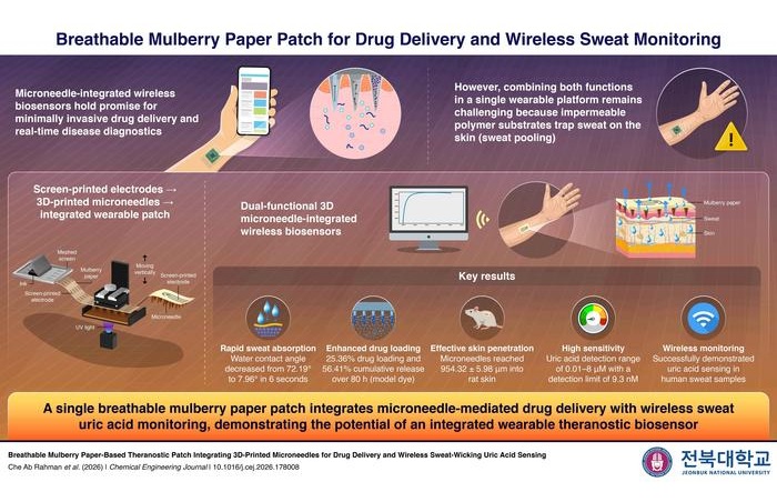
- Paper-Based Patch Integrates Sweat Sensors and Drug-Delivery Microneedles
- Circuit-Free Smart Patch Uses Smartphone Camera to Monitor Pulse and Vessel Stiffness
- Chest Digital Twin Guides Light Therapy Dosing for Pneumonia
- Intraoperative Drug Combination Reduces Kidney Injury in Septic Shock Surgery
- Wirelessly Powered Implant Targets Excess Glucose in the Blood
- FDA Approves First Transcatheter Device for Paravalvular Leak Closure
- Smaller Surgical Margins May Help Protect Healthy Tissue in Young Sarcoma Patients
- BD Launches TIPS Stent Graft for Personalized Portal Hypertension Treatment
- FDA-Cleared Thrombectomy System Removes Pulmonary Embolism Clots
- Implantable Ultrasound Device Enhances Local Drug Delivery in Glioblastoma
- Expert Review Outlines Diagnosis and Treatment of Recurrent Catheter Infections
- Total-Body PET/CT Scanner Enables One-Minute Diagnostic-Quality Cancer Imaging
- AI Platform Personalizes Support Between Visits for Cancer Survivors
- At-Home Neuromodulation Shows Promise in Selected Patients With Hypertension
- EHR Model Flags High-Risk Periods in Patients with Metastatic Breast Cancer
- Researchers Develop AI Tool to Reconstruct Missing Brain Connectivity Data
- Cardiac MRI AI Tool Improves Mortality Risk Prediction
- Imaging Biomarker Links Breast Tumor Heterogeneity to Targeted Therapy Response
- AI-Guided Echocardiography Enables Mobile Cardiac Imaging in Rural Communities
- Privacy-Preserving AI Tool Synthesizes CT for Radiation Therapy Planning
- Stryker Enters Soft-Tissue Robotics Through Strategic Alliance With CMR Surgical
- Terumo Neuro Acquisition Advances Liquid Embolic Technology for Chronic Subdural Hematoma
- World Health Expo to Showcase Trillion-Dollar Growth Across Pharma, Biotech, Longevity and Wellness at 2027 Edition
- Medtronic Receives FDA Clearance for Vessel-Sealing Instrument for Robotic Surgery
- Robotic Single-Port System Gains CE Mark for Transvaginal Gynecologic Procedures
- EHR Alerts Support Dosing, Sedation, and Shock Recognition in Cardiac Care
- Adaptive Radiation QA Software Gains Validation for NeoArc Treatments
- Healthcare Database Helps Close Long COVID Surveillance Gap
- New Tablet Platform Extends Stroke Neurorehabilitation Beyond the Clinic
- Smartphone App Improves Blood Pressure Control in Hypertension
- AI Tool Predicts Hypertensive Crisis Up to 72 Hours Ahead
- AI Tool Predicts Suicide Risk in Emergency Departments from Routine Data
- AI Chatbot Helps Women Identify Heart Disease Risks and Symptoms
- AI Tool Prioritizes Peer Referrals to Expand HIV Testing Access
- Explainable AI Dermoscopy Tool Supports Risk Assessment for Skin Cancer
- Continuous Glucose Monitoring Identifies Cardiometabolic Risk in Adults Without Diabetes
- Microneedle Patch Enables At-Home Monitoring of Acute Kidney Injury
- Ultrathin Metasurface Could Bring Quantitative Phase Imaging to Portable Devices
- Rapid Onsite Testing Reduces Emergency Department Transfers in Long-Term Care
- New Brain Ultrasound Platform Enables Bedside Postoperative Imaging
 Expo
- Paper-Based Patch Integrates Sweat Sensors and Drug-Delivery Microneedles
- Circuit-Free Smart Patch Uses Smartphone Camera to Monitor Pulse and Vessel Stiffness
- Chest Digital Twin Guides Light Therapy Dosing for Pneumonia
- Intraoperative Drug Combination Reduces Kidney Injury in Septic Shock Surgery
- Wirelessly Powered Implant Targets Excess Glucose in the Blood
- FDA Approves First Transcatheter Device for Paravalvular Leak Closure
- Smaller Surgical Margins May Help Protect Healthy Tissue in Young Sarcoma Patients
- BD Launches TIPS Stent Graft for Personalized Portal Hypertension Treatment
- FDA-Cleared Thrombectomy System Removes Pulmonary Embolism Clots
- Implantable Ultrasound Device Enhances Local Drug Delivery in Glioblastoma
- Expert Review Outlines Diagnosis and Treatment of Recurrent Catheter Infections
- Total-Body PET/CT Scanner Enables One-Minute Diagnostic-Quality Cancer Imaging
- AI Platform Personalizes Support Between Visits for Cancer Survivors
- At-Home Neuromodulation Shows Promise in Selected Patients With Hypertension
- EHR Model Flags High-Risk Periods in Patients with Metastatic Breast Cancer
- Researchers Develop AI Tool to Reconstruct Missing Brain Connectivity Data
- Cardiac MRI AI Tool Improves Mortality Risk Prediction
- Imaging Biomarker Links Breast Tumor Heterogeneity to Targeted Therapy Response
- AI-Guided Echocardiography Enables Mobile Cardiac Imaging in Rural Communities
- Privacy-Preserving AI Tool Synthesizes CT for Radiation Therapy Planning
- Stryker Enters Soft-Tissue Robotics Through Strategic Alliance With CMR Surgical
- Terumo Neuro Acquisition Advances Liquid Embolic Technology for Chronic Subdural Hematoma
- World Health Expo to Showcase Trillion-Dollar Growth Across Pharma, Biotech, Longevity and Wellness at 2027 Edition
- Medtronic Receives FDA Clearance for Vessel-Sealing Instrument for Robotic Surgery
- Robotic Single-Port System Gains CE Mark for Transvaginal Gynecologic Procedures
- EHR Alerts Support Dosing, Sedation, and Shock Recognition in Cardiac Care
- Adaptive Radiation QA Software Gains Validation for NeoArc Treatments
- Healthcare Database Helps Close Long COVID Surveillance Gap
- New Tablet Platform Extends Stroke Neurorehabilitation Beyond the Clinic
- Smartphone App Improves Blood Pressure Control in Hypertension
- AI Tool Predicts Hypertensive Crisis Up to 72 Hours Ahead
- AI Tool Predicts Suicide Risk in Emergency Departments from Routine Data
- AI Chatbot Helps Women Identify Heart Disease Risks and Symptoms
- AI Tool Prioritizes Peer Referrals to Expand HIV Testing Access
- Explainable AI Dermoscopy Tool Supports Risk Assessment for Skin Cancer
- Continuous Glucose Monitoring Identifies Cardiometabolic Risk in Adults Without Diabetes
- Microneedle Patch Enables At-Home Monitoring of Acute Kidney Injury
- Ultrathin Metasurface Could Bring Quantitative Phase Imaging to Portable Devices
- Rapid Onsite Testing Reduces Emergency Department Transfers in Long-Term Care
- New Brain Ultrasound Platform Enables Bedside Postoperative Imaging




































 Client Login