




Expo
view channel
view channel
view channel
view channel
view channel
view channel
view channel
view channel
AICritical CareSurgical TechniquesPatient Care
Health ITPoint of CareBusiness
Events
Webinars

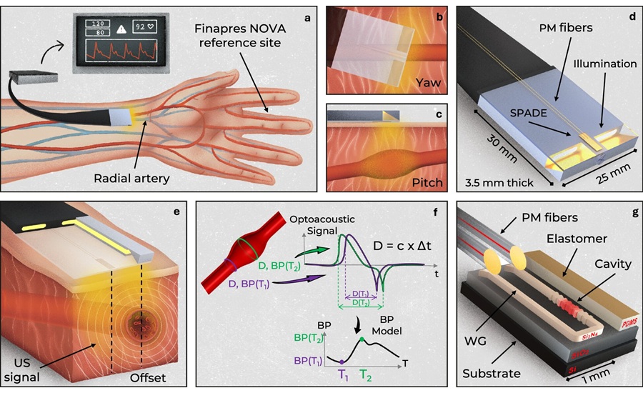
- Miniature Wearable Sensor Enables Continuous Cuffless Blood Pressure Monitoring
- Studies Support AI-ECG for Detecting Suspected Hypertrophic Cardiomyopathy
- Personalized fMRI-Guided TMS May Improve Severe OCD Symptoms
- Updated Staging System Standardizes Cardiogenic Shock Assessment
- Routine Opt-Out Screening Helps Increase Detection of Adolescent STIs
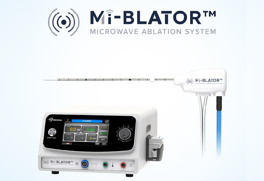
- Programmable Microwave Ablation System Gains CE Mark for Minimally Invasive Tissue Ablation
- Minimally Invasive Balloon Procedure May Redirect Blood Flow During Cardiac Arrest
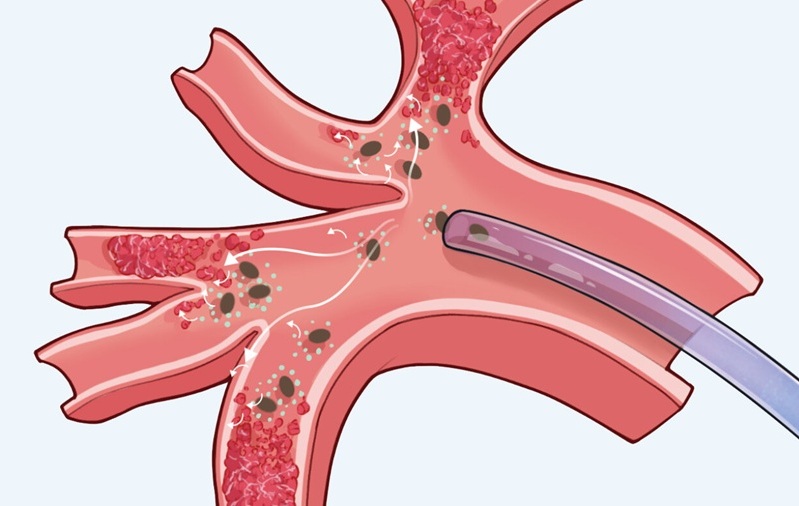
- Researchers Develop Microbots to Help Drugs Reach Blood Clots
- First Stemless Reverse Shoulder Arthroplasty System Receives FDA Clearance in U.S.
- First U.S. Procedures Successfully Completed With New GERD Implant
- Video-Based Support Helps Older Adults Clarify Kidney Disease Treatment Preferences
- Expert Review Outlines Diagnosis and Treatment of Recurrent Catheter Infections
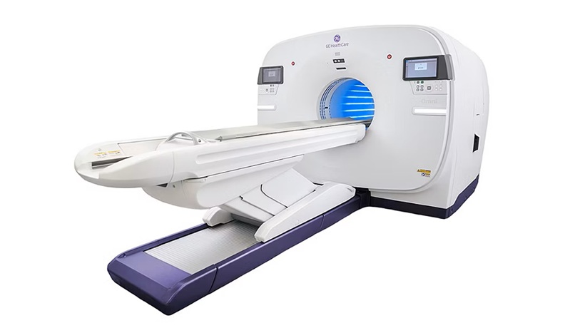
- Total-Body PET/CT Scanner Enables One-Minute Diagnostic-Quality Cancer Imaging
- AI Platform Personalizes Support Between Visits for Cancer Survivors
- At-Home Neuromodulation Shows Promise in Selected Patients With Hypertension
- MRI Test Identifies Candidates for Lower-Dose Radiotherapy in HPV Throat Cancer
- MRI-Guided Focused Ultrasound Improves Motor Symptoms in Parkinson’s Disease
- AI Tool Reconstructs Missing Brain Connectivity Data
- Targeted ImmunoPET Scan Detects Multiple Myeloma Missed by Standard PET/CT
- Cardiac MRI AI Tool Improves Mortality Risk Prediction
- Healthcare Partnership Creates Testbed for Advanced Connectivity and AI Applications
- Steris to Expand Surgical Safety Portfolio With $2.1 Billion Xodus Medical Deal
- Stryker Enters Soft-Tissue Robotics Through Strategic Alliance With CMR Surgical
- Terumo Neuro Acquisition Advances Liquid Embolic Technology for Chronic Subdural Hematoma
- World Health Expo to Showcase Trillion-Dollar Growth Across Pharma, Biotech, Longevity and Wellness at 2027 Edition
- EHR Alerts Support Dosing, Sedation, and Shock Recognition in Cardiac Care
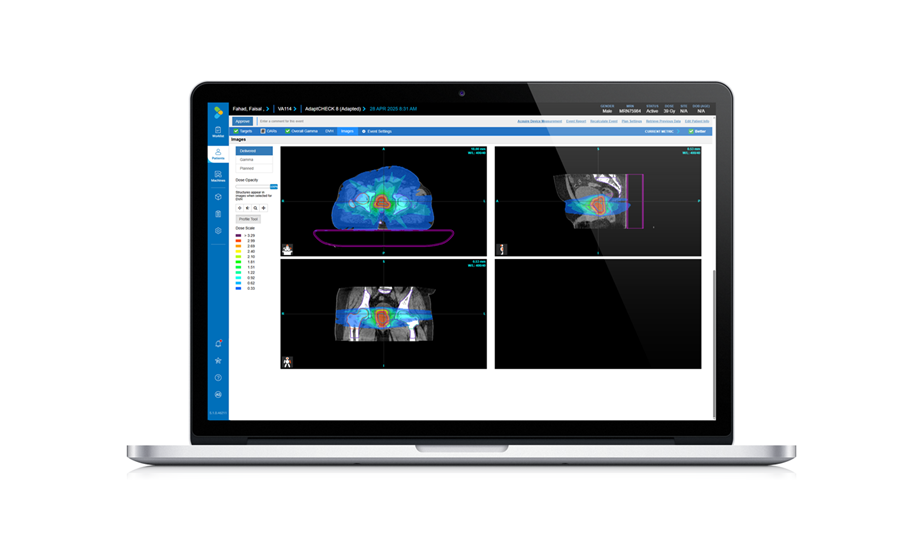
- Adaptive Radiation QA Software Gains Validation for NeoArc Treatments
- Healthcare Database Helps Close Long COVID Surveillance Gap
- New Tablet Platform Extends Stroke Neurorehabilitation Beyond the Clinic
- Smartphone App Improves Blood Pressure Control in Hypertension
- AI Tool Speeds Echocardiogram Reporting With Comparable Accuracy
- AI Tool Improves Hospital Discharge Summaries to Support Follow-Up Care
- AI Tool Predicts Hypertensive Crisis Up to 72 Hours Ahead
- AI Tool Predicts Suicide Risk in Emergency Departments from Routine Data
- AI Chatbot Helps Women Identify Heart Disease Risks and Symptoms
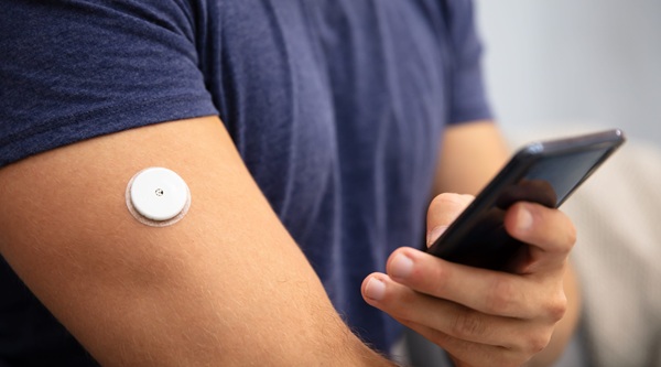
- Continuous Glucose Monitoring Identifies Cardiometabolic Risk in Adults Without Diabetes
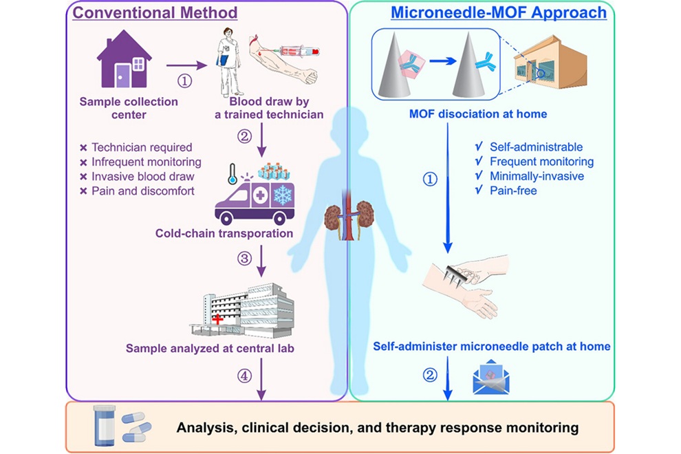
- Microneedle Patch Enables At-Home Monitoring of Acute Kidney Injury
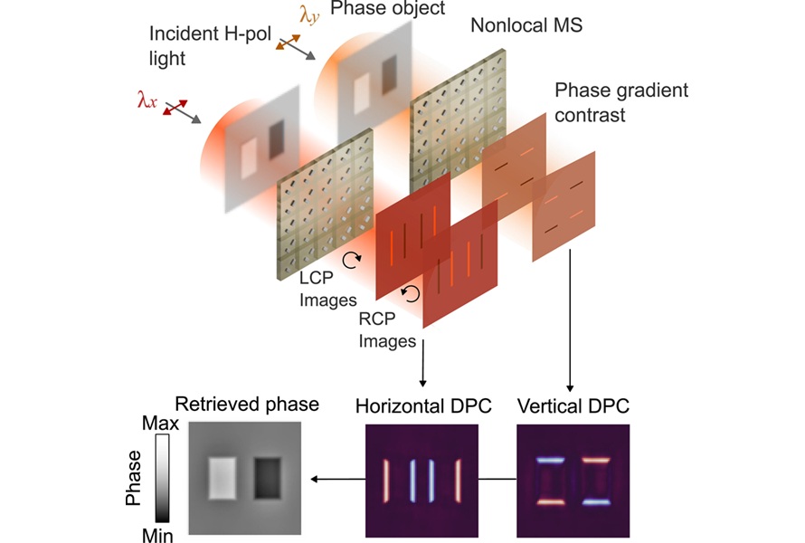
- Ultrathin Metasurface Could Bring Quantitative Phase Imaging to Portable Devices
- Rapid Onsite Testing Reduces Emergency Department Transfers in Long-Term Care
- New Brain Ultrasound Platform Enables Bedside Postoperative Imaging
 Expo
- Miniature Wearable Sensor Enables Continuous Cuffless Blood Pressure Monitoring
- Studies Support AI-ECG for Detecting Suspected Hypertrophic Cardiomyopathy
- Personalized fMRI-Guided TMS May Improve Severe OCD Symptoms
- Updated Staging System Standardizes Cardiogenic Shock Assessment
- Routine Opt-Out Screening Helps Increase Detection of Adolescent STIs
- Programmable Microwave Ablation System Gains CE Mark for Minimally Invasive Tissue Ablation
- Minimally Invasive Balloon Procedure May Redirect Blood Flow During Cardiac Arrest
- Researchers Develop Microbots to Help Drugs Reach Blood Clots
- First Stemless Reverse Shoulder Arthroplasty System Receives FDA Clearance in U.S.
- First U.S. Procedures Successfully Completed With New GERD Implant
- Video-Based Support Helps Older Adults Clarify Kidney Disease Treatment Preferences
- Expert Review Outlines Diagnosis and Treatment of Recurrent Catheter Infections
- Total-Body PET/CT Scanner Enables One-Minute Diagnostic-Quality Cancer Imaging
- AI Platform Personalizes Support Between Visits for Cancer Survivors
- At-Home Neuromodulation Shows Promise in Selected Patients With Hypertension
- MRI Test Identifies Candidates for Lower-Dose Radiotherapy in HPV Throat Cancer
- MRI-Guided Focused Ultrasound Improves Motor Symptoms in Parkinson’s Disease
- AI Tool Reconstructs Missing Brain Connectivity Data
- Targeted ImmunoPET Scan Detects Multiple Myeloma Missed by Standard PET/CT
- Cardiac MRI AI Tool Improves Mortality Risk Prediction
- Healthcare Partnership Creates Testbed for Advanced Connectivity and AI Applications
- Steris to Expand Surgical Safety Portfolio With $2.1 Billion Xodus Medical Deal
- Stryker Enters Soft-Tissue Robotics Through Strategic Alliance With CMR Surgical
- Terumo Neuro Acquisition Advances Liquid Embolic Technology for Chronic Subdural Hematoma
- World Health Expo to Showcase Trillion-Dollar Growth Across Pharma, Biotech, Longevity and Wellness at 2027 Edition
- EHR Alerts Support Dosing, Sedation, and Shock Recognition in Cardiac Care
- Adaptive Radiation QA Software Gains Validation for NeoArc Treatments
- Healthcare Database Helps Close Long COVID Surveillance Gap
- New Tablet Platform Extends Stroke Neurorehabilitation Beyond the Clinic
- Smartphone App Improves Blood Pressure Control in Hypertension
- AI Tool Speeds Echocardiogram Reporting With Comparable Accuracy
- AI Tool Improves Hospital Discharge Summaries to Support Follow-Up Care
- AI Tool Predicts Hypertensive Crisis Up to 72 Hours Ahead
- AI Tool Predicts Suicide Risk in Emergency Departments from Routine Data
- AI Chatbot Helps Women Identify Heart Disease Risks and Symptoms
- Continuous Glucose Monitoring Identifies Cardiometabolic Risk in Adults Without Diabetes
- Microneedle Patch Enables At-Home Monitoring of Acute Kidney Injury
- Ultrathin Metasurface Could Bring Quantitative Phase Imaging to Portable Devices
- Rapid Onsite Testing Reduces Emergency Department Transfers in Long-Term Care
- New Brain Ultrasound Platform Enables Bedside Postoperative Imaging




























 Client Login