




Expo
view channel
view channel
view channel
view channel
view channel
view channel
view channel
view channel
AICritical CareSurgical TechniquesPatient Care
Health ITPoint of CareBusiness
Events
Webinars

- Real-Time Data Platform Supports Precision Care in Polycystic Kidney Disease
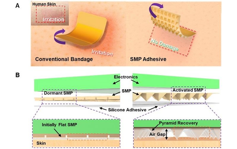
- Smart Medical Adhesive Patch Uses Heat to Enable Gentle Removal
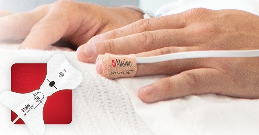
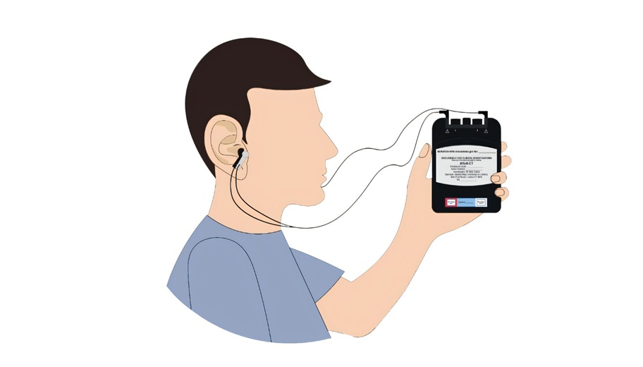
- Smart Pulse Oximetry Supports Earlier Recognition of Arrhythmias and Respiratory Risk
- Proposed Risk Score Prioritizes Sickest Patients for Heart Transplant
- Wearable Robotic Gait Training Linked to Improved Walking Distance in ALS
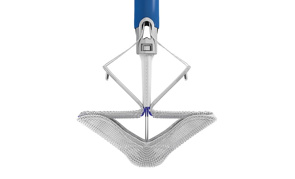
- Transcatheter Repair System Gains FDA Approval for Severe Tricuspid Regurgitation
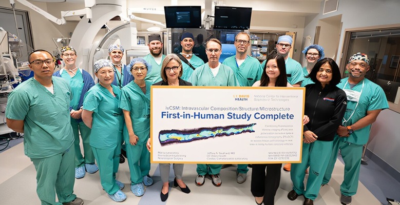
- Dual-Imaging Catheter Completes Initial Human Evaluation of Coronary Plaque
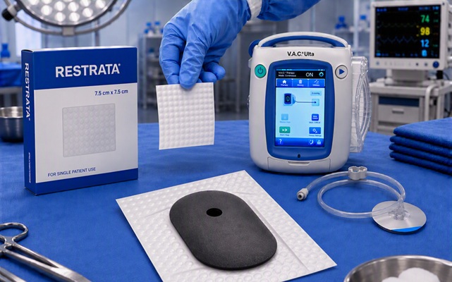
- Combined Wound Healing Approach Pairs Synthetic Scaffold With Negative Pressure Therapy
- Shape-Changing Hydrogels Show Promise for Tissue Engineering and Wound Healing
- Augmented Reality Helps Locate Hidden Tumors During Liver Surgery
- AI Platform Personalizes Support Between Visits for Cancer Survivors
- At-Home Neuromodulation Shows Promise in Selected Patients With Hypertension
- EHR Model Flags High-Risk Periods in Patients with Metastatic Breast Cancer
- Virtual Reality Helps Claustrophobic Patients Complete Cardiac PET/CT Scans
- Weighted Blankets Reduce Anxiety in Hospitalized Adults
- AI Tool Improves Fetal MRI for Brain Abnormality Diagnosis
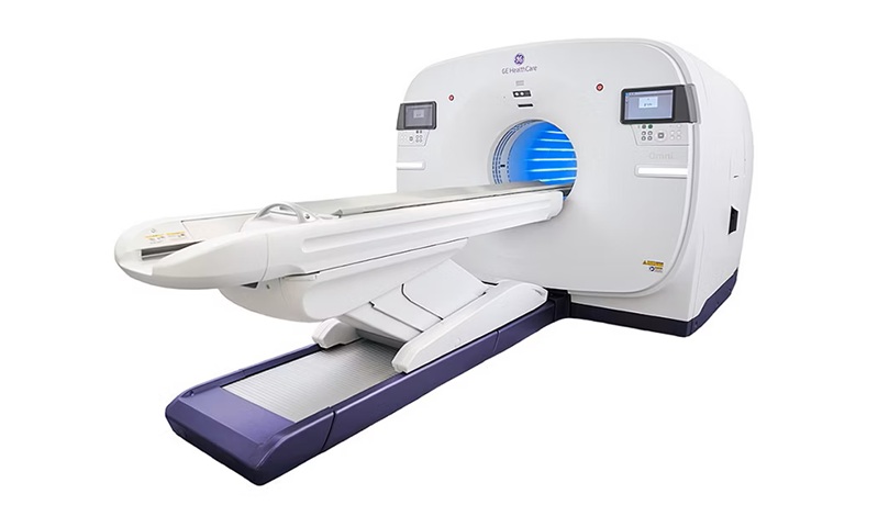
- AI Model Detects Hidden Disease Signals in Routine CT Scans
- Targeted PET/CT Predicts Early Treatment Response in HER2-Positive Breast Cancer
- Amyloid and Tau PET Guide Selection and Monitoring of Alzheimer’s Therapy
- MRI Tool Analysis Identifies Vascular Differences in Cerebral Small Vessel Disease
- Stryker Enters Soft-Tissue Robotics Through Strategic Alliance With CMR Surgical
- Terumo Neuro Acquisition Advances Liquid Embolic Technology for Chronic Subdural Hematoma
- World Health Expo to Showcase Trillion-Dollar Growth Across Pharma, Biotech, Longevity and Wellness at 2027 Edition
- Medtronic Receives FDA Clearance for Vessel-Sealing Instrument for Robotic Surgery
- Robotic Single-Port System Gains CE Mark for Transvaginal Gynecologic Procedures
- EHR Alerts Support Dosing, Sedation, and Shock Recognition in Cardiac Care
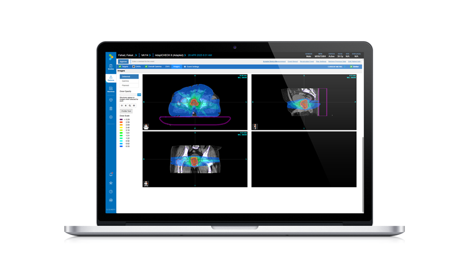
- Adaptive Radiation QA Software Gains Validation for NeoArc Treatments
- Healthcare Database Helps Close Long COVID Surveillance Gap
- New Tablet Platform Extends Stroke Neurorehabilitation Beyond the Clinic
- Smartphone App Improves Blood Pressure Control in Hypertension
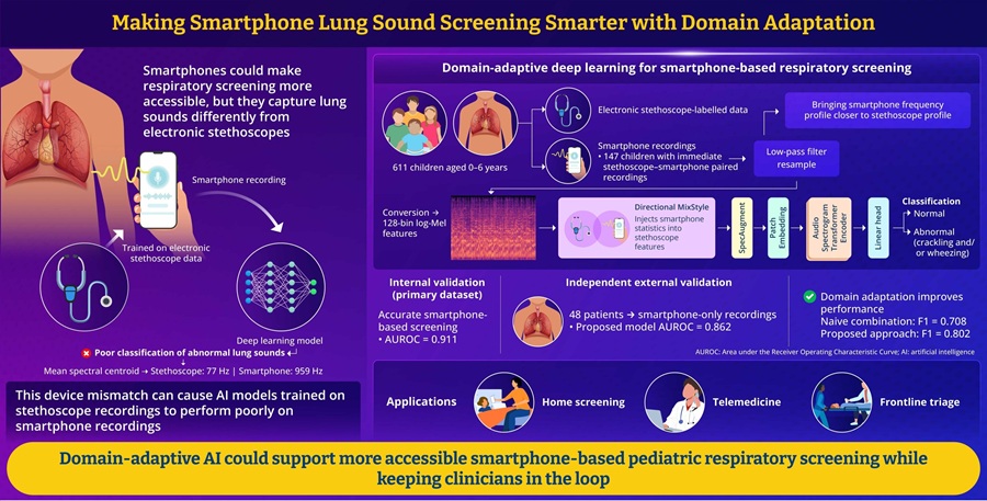
- AI Tool Improves Smartphone Detection of Abnormal Pediatric Lung Sounds
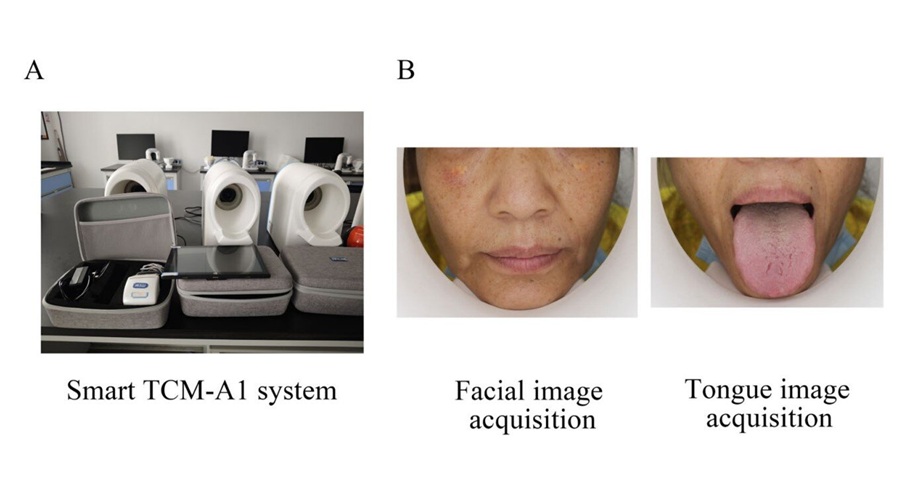
- AI Tool Detects Coronary Heart Disease from Face and Tongue Images
- Optical Imaging System Helps Predict Chemotherapy Response in Cancer
- AI Tool Predicts Long-Term Cardiovascular Risk from Sleep Study ECG
- Predictive Model Supports Growth Hormone Treatment Planning in Idiopathic Short Stature
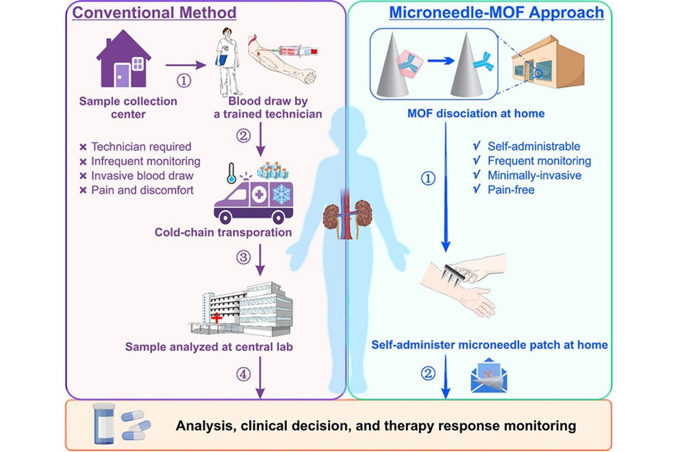
- Microneedle Patch Enables At-Home Monitoring of Acute Kidney Injury
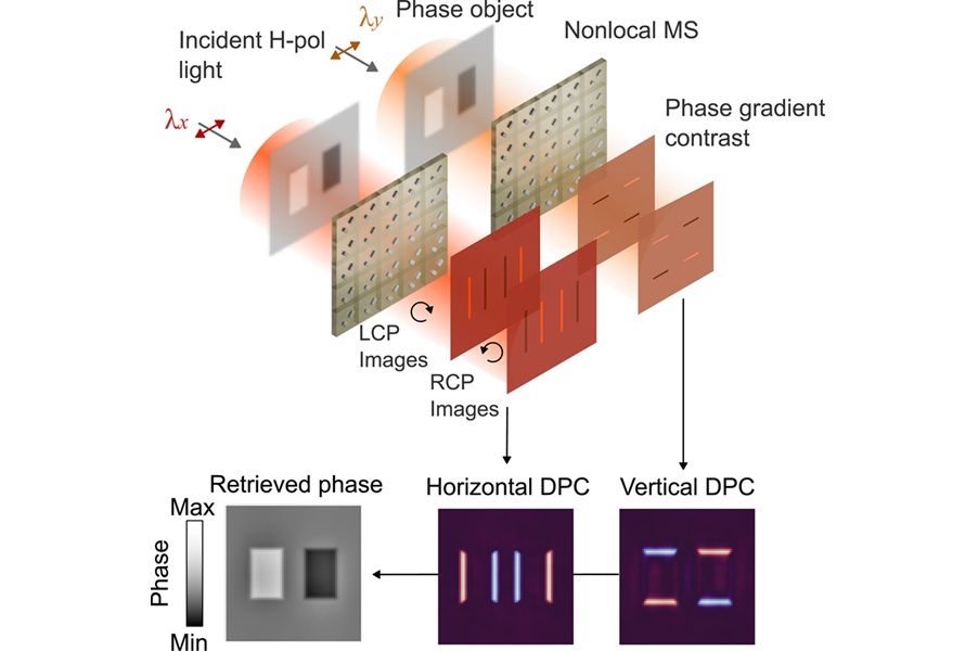
- Ultrathin Metasurface Could Bring Quantitative Phase Imaging to Portable Devices
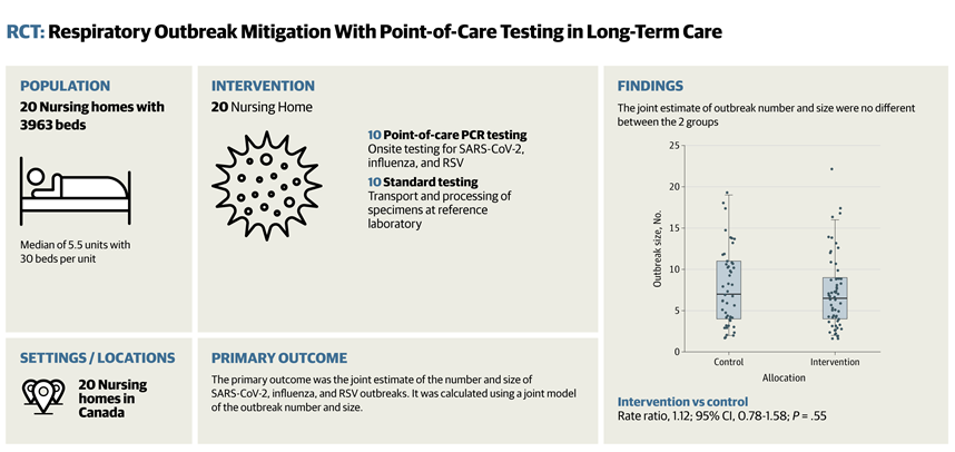
- Rapid Onsite Testing Reduces Emergency Department Transfers in Long-Term Care
- New Brain Ultrasound Platform Enables Bedside Postoperative Imaging
- Handheld Ultrasound Expands Point-of-Care Imaging Access in Brazil
 Expo
Expo
- Real-Time Data Platform Supports Precision Care in Polycystic Kidney Disease
- Smart Medical Adhesive Patch Uses Heat to Enable Gentle Removal
- Smart Pulse Oximetry Supports Earlier Recognition of Arrhythmias and Respiratory Risk
- Proposed Risk Score Prioritizes Sickest Patients for Heart Transplant
- Wearable Robotic Gait Training Linked to Improved Walking Distance in ALS
- Transcatheter Repair System Gains FDA Approval for Severe Tricuspid Regurgitation
- Dual-Imaging Catheter Completes Initial Human Evaluation of Coronary Plaque
- Combined Wound Healing Approach Pairs Synthetic Scaffold With Negative Pressure Therapy
- Shape-Changing Hydrogels Show Promise for Tissue Engineering and Wound Healing
- Augmented Reality Helps Locate Hidden Tumors During Liver Surgery
- AI Platform Personalizes Support Between Visits for Cancer Survivors
- At-Home Neuromodulation Shows Promise in Selected Patients With Hypertension
- EHR Model Flags High-Risk Periods in Patients with Metastatic Breast Cancer
- Virtual Reality Helps Claustrophobic Patients Complete Cardiac PET/CT Scans
- Weighted Blankets Reduce Anxiety in Hospitalized Adults
- AI Tool Improves Fetal MRI for Brain Abnormality Diagnosis
- AI Model Detects Hidden Disease Signals in Routine CT Scans
- Targeted PET/CT Predicts Early Treatment Response in HER2-Positive Breast Cancer
- Amyloid and Tau PET Guide Selection and Monitoring of Alzheimer’s Therapy
- MRI Tool Analysis Identifies Vascular Differences in Cerebral Small Vessel Disease
- Stryker Enters Soft-Tissue Robotics Through Strategic Alliance With CMR Surgical
- Terumo Neuro Acquisition Advances Liquid Embolic Technology for Chronic Subdural Hematoma
- World Health Expo to Showcase Trillion-Dollar Growth Across Pharma, Biotech, Longevity and Wellness at 2027 Edition
- Medtronic Receives FDA Clearance for Vessel-Sealing Instrument for Robotic Surgery
- Robotic Single-Port System Gains CE Mark for Transvaginal Gynecologic Procedures
- EHR Alerts Support Dosing, Sedation, and Shock Recognition in Cardiac Care
- Adaptive Radiation QA Software Gains Validation for NeoArc Treatments
- Healthcare Database Helps Close Long COVID Surveillance Gap
- New Tablet Platform Extends Stroke Neurorehabilitation Beyond the Clinic
- Smartphone App Improves Blood Pressure Control in Hypertension
- AI Tool Improves Smartphone Detection of Abnormal Pediatric Lung Sounds
- AI Tool Detects Coronary Heart Disease from Face and Tongue Images
- Optical Imaging System Helps Predict Chemotherapy Response in Cancer
- AI Tool Predicts Long-Term Cardiovascular Risk from Sleep Study ECG
- Predictive Model Supports Growth Hormone Treatment Planning in Idiopathic Short Stature
- Microneedle Patch Enables At-Home Monitoring of Acute Kidney Injury
- Ultrathin Metasurface Could Bring Quantitative Phase Imaging to Portable Devices
- Rapid Onsite Testing Reduces Emergency Department Transfers in Long-Term Care
- New Brain Ultrasound Platform Enables Bedside Postoperative Imaging
- Handheld Ultrasound Expands Point-of-Care Imaging Access in Brazil





























 Client Login