




Expo
Medica
view channel
view channel
view channel
view channel
view channel
view channel
view channel
view channel
Critical CareSurgical TechniquesPatient CareMedical ImagingHealth ITPoint of CareBusiness
Events
Webinars

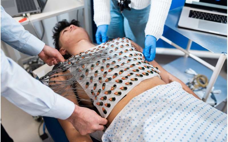
- Wearable Heart Mapping May Help Identify Cardiovascular Hospitalization Risk
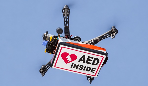
- Drone Network Could Speed AED Delivery for Out-of-Hospital Cardiac Arrest
- AI Models Improve Mortality Risk Prediction After PCI for STEMI
- Updated Head Injury Guidelines Support Risk-Based CT and Observation Decisions
- AI-Derived Heart Age Linked to Cardiovascular and Dementia-Related Mortality
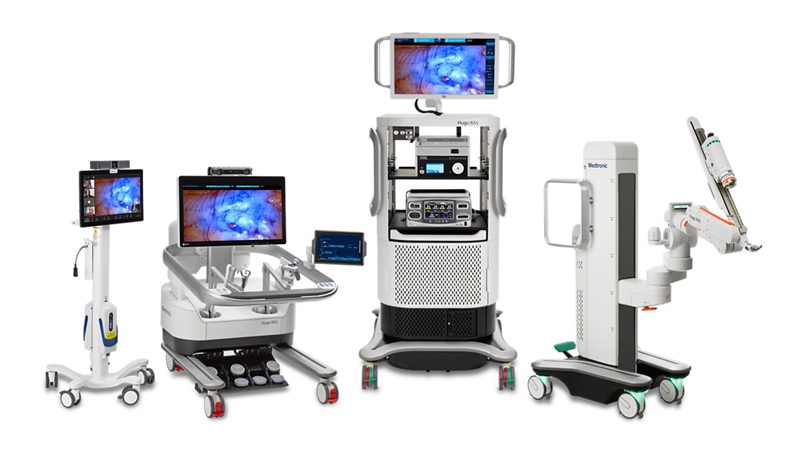
- FDA Authorizes Robotic System for En Bloc Bladder Resection
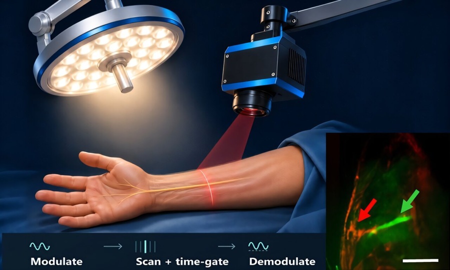
- Fluorescence Imaging Reveals Nerves Under Normal Operating-Room Lighting
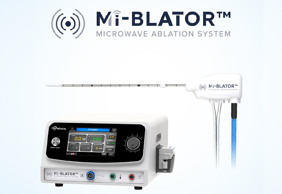
- Programmable Microwave Ablation System Gains CE Mark for Minimally Invasive Tissue Ablation
- Minimally Invasive Balloon Procedure May Redirect Blood Flow During Cardiac Arrest
- Researchers Develop Microbots to Help Drugs Reach Blood Clots
- Video-Based Support Helps Older Adults Clarify Kidney Disease Treatment Preferences
- Expert Review Outlines Diagnosis and Treatment of Recurrent Catheter Infections
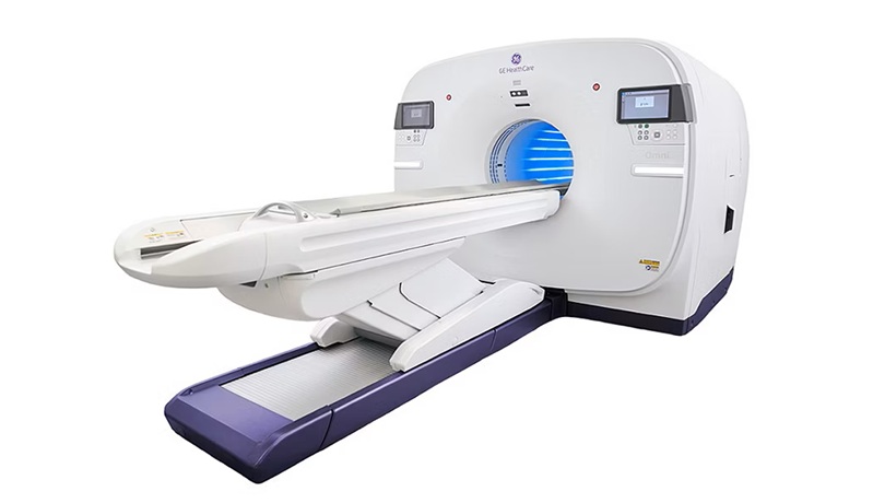
- Total-Body PET/CT Scanner Enables One-Minute Diagnostic-Quality Cancer Imaging
- AI Platform Personalizes Support Between Visits for Cancer Survivors
- At-Home Neuromodulation Shows Promise in Selected Patients With Hypertension
- AI Combines Brain Imaging Features to Identify Cognitive Impairment in CKD
- AI Software Gains FDA Clearance for Brain Aneurysm Detection and Follow-Up
- Lung Ultrasound Predicts Progression of Interstitial Lung Abnormalities in Rheumatoid Arthritis
- MRI Test Identifies Candidates for Lower-Dose Radiotherapy in HPV Throat Cancer
- MRI-Guided Focused Ultrasound Improves Motor Symptoms in Parkinson’s Disease
- Healthcare Partnership Creates Testbed for Advanced Connectivity and AI Applications
- Steris to Expand Surgical Safety Portfolio With $2.1 Billion Xodus Medical Deal
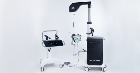
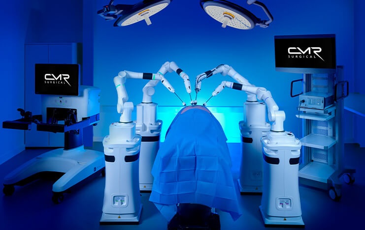
- Stryker Enters Soft-Tissue Robotics Through Strategic Alliance With CMR Surgical
- Terumo Neuro Acquisition Advances Liquid Embolic Technology for Chronic Subdural Hematoma
- World Health Expo to Showcase Trillion-Dollar Growth Across Pharma, Biotech, Longevity and Wellness at 2027 Edition
- Clinical Decision Support Tool Automates CKD Risk Assessment in Primary Care
- EHR Alerts Support Dosing, Sedation, and Shock Recognition in Cardiac Care
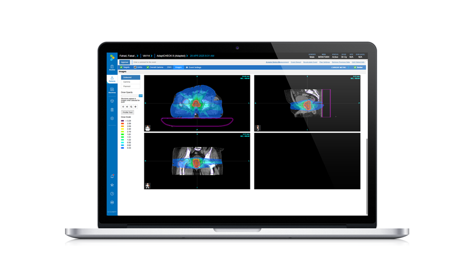
- Adaptive Radiation QA Software Gains Validation for NeoArc Treatments
- Healthcare Database Helps Close Long COVID Surveillance Gap
- New Tablet Platform Extends Stroke Neurorehabilitation Beyond the Clinic
- Causal AI Tool Withholds Ovarian Cancer Treatment Recommendations When Evidence Insufficient
- AI Analysis of Smartwatch ECGs May Help Detect Weakened Heart Function
- AI Tool Speeds Echocardiogram Reporting With Comparable Accuracy
- AI Tool Improves Hospital Discharge Summaries to Support Follow-Up Care
- AI Tool Predicts Hypertensive Crisis Up to 72 Hours Ahead
- New Breath Condensate Testing Approach Enhances Reliability for COPD Monitoring
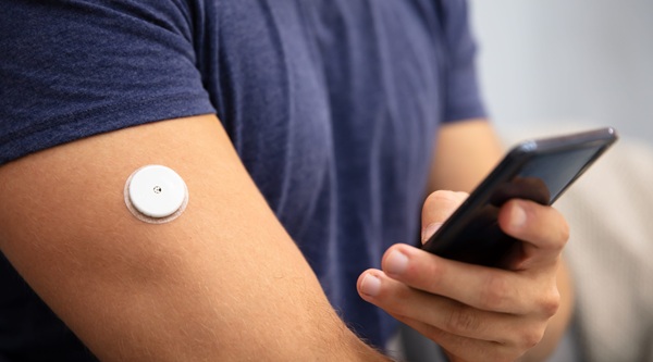
- Continuous Glucose Monitoring Identifies Cardiometabolic Risk in Adults Without Diabetes
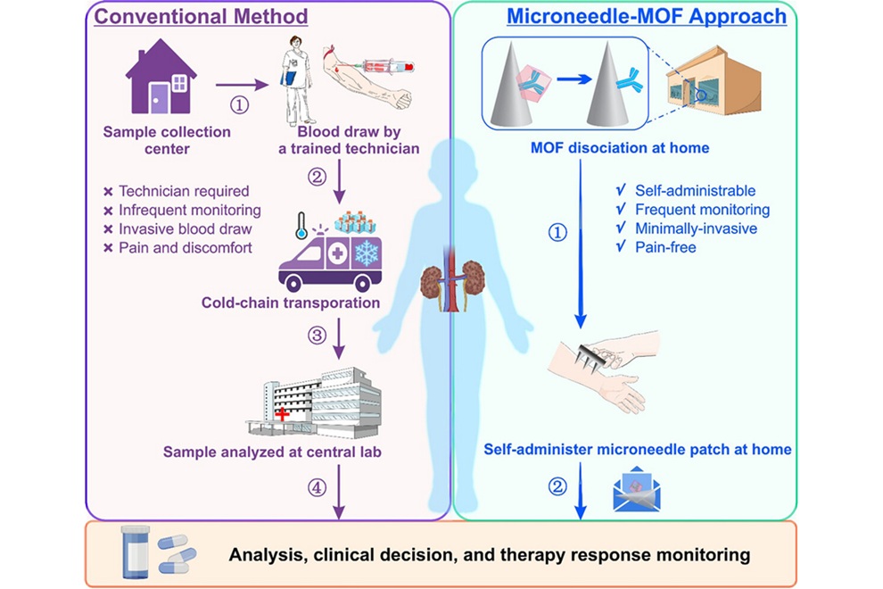
- Microneedle Patch Enables At-Home Monitoring of Acute Kidney Injury
- Ultrathin Metasurface Could Bring Quantitative Phase Imaging to Portable Devices
- Rapid Onsite Testing Reduces Emergency Department Transfers in Long-Term Care
 Expo
Medica
- Wearable Heart Mapping May Help Identify Cardiovascular Hospitalization Risk
- Drone Network Could Speed AED Delivery for Out-of-Hospital Cardiac Arrest
- AI Models Improve Mortality Risk Prediction After PCI for STEMI
- Updated Head Injury Guidelines Support Risk-Based CT and Observation Decisions
- AI-Derived Heart Age Linked to Cardiovascular and Dementia-Related Mortality
- FDA Authorizes Robotic System for En Bloc Bladder Resection
- Fluorescence Imaging Reveals Nerves Under Normal Operating-Room Lighting
- Programmable Microwave Ablation System Gains CE Mark for Minimally Invasive Tissue Ablation
- Minimally Invasive Balloon Procedure May Redirect Blood Flow During Cardiac Arrest
- Researchers Develop Microbots to Help Drugs Reach Blood Clots
- Video-Based Support Helps Older Adults Clarify Kidney Disease Treatment Preferences
- Expert Review Outlines Diagnosis and Treatment of Recurrent Catheter Infections
- Total-Body PET/CT Scanner Enables One-Minute Diagnostic-Quality Cancer Imaging
- AI Platform Personalizes Support Between Visits for Cancer Survivors
- At-Home Neuromodulation Shows Promise in Selected Patients With Hypertension
- AI Combines Brain Imaging Features to Identify Cognitive Impairment in CKD
- AI Software Gains FDA Clearance for Brain Aneurysm Detection and Follow-Up
- Lung Ultrasound Predicts Progression of Interstitial Lung Abnormalities in Rheumatoid Arthritis
- MRI Test Identifies Candidates for Lower-Dose Radiotherapy in HPV Throat Cancer
- MRI-Guided Focused Ultrasound Improves Motor Symptoms in Parkinson’s Disease
- Healthcare Partnership Creates Testbed for Advanced Connectivity and AI Applications
- Steris to Expand Surgical Safety Portfolio With $2.1 Billion Xodus Medical Deal
- Stryker Enters Soft-Tissue Robotics Through Strategic Alliance With CMR Surgical
- Terumo Neuro Acquisition Advances Liquid Embolic Technology for Chronic Subdural Hematoma
- World Health Expo to Showcase Trillion-Dollar Growth Across Pharma, Biotech, Longevity and Wellness at 2027 Edition
- Clinical Decision Support Tool Automates CKD Risk Assessment in Primary Care
- EHR Alerts Support Dosing, Sedation, and Shock Recognition in Cardiac Care
- Adaptive Radiation QA Software Gains Validation for NeoArc Treatments
- Healthcare Database Helps Close Long COVID Surveillance Gap
- New Tablet Platform Extends Stroke Neurorehabilitation Beyond the Clinic
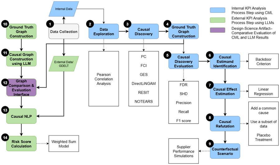
- Causal AI Tool Withholds Ovarian Cancer Treatment Recommendations When Evidence Insufficient
- AI Analysis of Smartwatch ECGs May Help Detect Weakened Heart Function
- AI Tool Speeds Echocardiogram Reporting With Comparable Accuracy
- AI Tool Improves Hospital Discharge Summaries to Support Follow-Up Care
- AI Tool Predicts Hypertensive Crisis Up to 72 Hours Ahead
- Richard Wolf to Present Innovative System Solutions at Germany Trade Show
- PROTEC to Launch New Motorized PRS 500 B X-ray System
- Randox to Showcase Future-Proofing Diagnostic Technology at Trade Fair
- NDS Announces New OR Networking Solution at Germany Trade Show
- Global Good and Motic Announce AI-Powered Microscope at MEDICA 2017
- Rober Showcases Pioneering ‘Zero Pressure’ Mattresses in Germany
- UK Companies Showcase Life-Saving Solutions at Medica 2018
- Siemens Healthineers Showcases ACUSON Ultrasound Product Portfolio
- Metaltronica Presents Digital Mammography Systems at Medical Trade Fair
- Canon Displays New Products at MEDICA Show
- Samsung Medison Exhibits New HERA I10 Combination Ultrasound System
- Medtronic Displays Patient Monitoring Systems at MEDICA 2019
- Healcerion Exhibits SONON Wireless Handheld Ultrasound at MEDICA 2019
- Hans Rudolph Exhibits Latest Range of Oro-Nasal Masks at MEDICA 2019
- EIZO Demonstrates CuratOR Alipe IP-Based Video Management Solution at MEDICA 2019
- Fisher & Paykel Demonstrates Humidified Nasal High Flow System for Delivering Respiratory Support at MEDICA 2021
- Axcent Medical Showcases Premium ICU Ventilator and Electronic Anesthesia Workstation at MEDICA 2021
- MESI Demonstrates Revolutionary mTABLET System at MEDICA 2021
- Microlife Presents the Only Blood Pressure Monitor with Integrated AFIB Detection at MEDICA 2021
- Innovative Health Presents Its Breakthrough Versatile and Low Cost Non-Electric Infusion System at MEDICA 2021
- Huntleigh Exhibits Range of Vital Signs Monitoring Solutions at MEDICA 2022
- Owen Mumford Presents Cutting-Edge Capillary Blood Sampling Devices for POC Testing
- Noul Demonstrates AI-Driven On-Site Diagnostics Platform at MEDICA 2022
- KUKA Presents Robot-Based Assistance Systems at MEDICA 2022
- Heyer Medical Exhibits State-Of-The-Art Ventilators, Anesthesia Workstations and Surgical Lights
- New Breath Condensate Testing Approach Enhances Reliability for COPD Monitoring
- Continuous Glucose Monitoring Identifies Cardiometabolic Risk in Adults Without Diabetes
- Microneedle Patch Enables At-Home Monitoring of Acute Kidney Injury
- Ultrathin Metasurface Could Bring Quantitative Phase Imaging to Portable Devices
- Rapid Onsite Testing Reduces Emergency Department Transfers in Long-Term Care
- LG Electronics Exhibits High Resolution Monitors for Surgery and Medical Diagnosis
- Advantech Demonstrates High-Performance Healthcare Systems and Solutions
- MAVIG Highlights X-Ray Protective Clothing and Suspension Systems for Medical Equipment
- EIZO GmbH Presents New 4K Visualization Solutions for Operating Room
- OptoMedic Showcases World of Fluorescence Endoscopy Technologies
- Norav Medical Demonstrates Advanced ECG Solutions
- MAVIG Highlights Latest Innovations in X-Ray Protection and Medical Suspension Systems
- EndoSemio Features Wireless Endoscope Tracheostomy Kit for Advanced Minimally Invasive Procedures
- Brain Navi Demonstrates First Autonomous Surgical Navigation Robot for Neurosurgery
- Winners Announced of the 14th MEDICA Start-up Competition
- Hurotics Demonstrates Advanced Gait Rehabilitation Exosuit at MEDICA 2025
- CETA Testsysteme Brings Advanced Leak and Flow Testing Technologies to MEDICA 2025
- Surgical Holdings Highlights Growth and Collaboration as Part of the Stille Group at MEDICA 2025
- WEGO Medical Brings Advanced Blood Management and Surgical Technologies to MEDICA 2025
- Winners Announced of the 15th MEDICA Start-up Competition
- EDAN Presents Medical Imaging, Patient Monitoring, and Diagnostic Solutions
- Hope Care and Huawei Showcase Remote Patient Monitoring Solutions at MEDICA 2026
- Taiwan Flagship Showcase Brings Medical Innovations Together at MEDICA 2026
- Transtek Presents Remote Patient Monitoring Devices and Connected Care Solutions































 Client Login