We use cookies to understand how you use our site and to improve your experience. This includes personalizing content and advertising. To learn more, click here. By continuing to use our site, you accept our use of cookies. Cookie Policy.

Features Partner Sites Information LinkXpress hp
Sign In
Advertise with Us
RANDOX LABORATORIES

Download Mobile App


3D-Printed Scaffolds Reconstruct Craniofacial Defects

By HospiMedica International staff writers
Posted on 03 Apr 2018
Print article
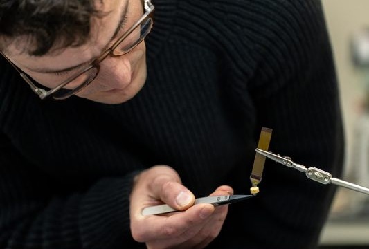
Image: Dr. Venu Varanasi and Research Assistant Tugba Cebe set the coordinates for the 3D printer (Photo courtesy of TAMU).
In-situ three-dimensional (3D) printing of osteogenic (bone regenerating) scaffolds can be used for the proper and rapid healing of bone fractures, claims a new study.

Developed by researchers at Texas A&M University (TAMU; College Station, TX, USA), the University of Texas (Arlington, USA), and other institutions, the substrate ink for the biosilica-biopolymer scaffold was prepared by mixing Laponite (Lp) with methacrylated gelatin (MAG); sucrose was used to increase viscosity and reduce gelation of the printing ink. During additive printing, crosslinking was initiated by ultra-violet (UV) light at the tip of the printer nozzle and, and the scaffolds were 3D printed in-situ, directly into calvaria bone defects using varied Laponite concentration so as to determine optimal bone density and chemical structure.

The scaffolds were fabricated into a mesh design, with dimensions matching that of formed defects; after four weeks, cranial bone samples were extracted. Evaluation by micro-CT showed that nearly 55% of the bone defect was healed for higher Lp- rich-MAG scaffolds, whereas empty control defects only had 11% of the defect filled with bone after four weeks. Histological staining showed that the scaffolds recruited osteoblasts and blood and growth factors into their structure to regenerate the intra-bony layers needed to initiate the healing process. The study was presented at the International & American Associations for Dental Research annual meeting, held during March 2018 in Fort Lauderdale (FL, USA).

“The results showed that 3D in-situ printing of bone regenerating scaffolds did improve the delivery of regenerative and reconstructive biomedical devices for the proper and rapid healing of bone fractures,” said senior author and study presenter Venu Varanasi, PhD, of TAMU. “This provides an advantage in that cells from within the initial hematoma become incorporated into the scaffold structure, thus, giving the operator flexibility to use the printed scaffold as a structural support that stimulates healing.”

“The gold standard for reconstruction of craniofacial defects involves carving of the cranial bone, hip bone, or the leg bone to recreate the missing structures. This is technically impossible for large facial defects,” said maxillofacial surgeon Likith Reddy, DDS, MD, director of residency training at the TAMU School of Dentistry. “If the technology works as anticipated, it will revolutionize the reconstruction of such complex three-dimensional structures. Reconstruction of such complex facial bony defects would become less of an art and more of a science.”

Severe traumatic injuries to the cranium have been challenging to heal due to the large missing bone volume. Typically, metal or plastic implants are used. But, these implants can take a long time to be customized for fit and often take a longer than desired time to support bone fixation. This can often lead to multiple revision surgeries if the defect is not properly healed. Moreover, the tissue that adjoins the implant can improperly heal.

Related Links:
Texas A&M University
University of Texas
Gold Member
Real-Time Diagnostics Onscreen Viewer
GEMweb Live
Flocked Fiber Swabs
Puritan® patented HydraFlock®
New
Mobile Autoclave
3870 HSG
New
Mammography Analytics Platform
Unifi Analytics Software

Print article
Radcal

Channels

Critical Care

view channel
Image: Overview concept and material design of the bioabsorbable electrical stimulation suture (BioES-suture) for treating muscle gashes (Photo courtesy of Nature Communications; DOI: 10.1038/s41467-024-52354-x)

Cutting-Edge Intelligent Medical Sutures Accelerate Wound Healing

In surgical medicine, sutures are the standard treatment for large incisions, but traditional sutures have functional limitations. Electrical stimulation is a non-drug therapy known to enhance wound healing.... Read more

Surgical Techniques

view channel
Image: Fluorescein-enhanced contrast imaging shows a rabbit’s normal sciatic nerve, left, and a damaged one (Photo courtesy of Osaka Metropolitan University)

Glowing Approach Helps Surgeons Assess Neural Blood Flow in Chronic Nerve Compression Neuropathy

In today's office environment, preventing the onset of carpal tunnel syndrome can be a daily challenge. In severe cases, surgery may be required to relieve nerve compression or repair damaged nerves.... Read more

Patient Care

view channel
Image: The portable, handheld BeamClean technology inactivates pathogens on commonly touched surfaces in seconds (Photo courtesy of Freestyle Partners)

First-Of-Its-Kind Portable Germicidal Light Technology Disinfects High-Touch Clinical Surfaces in Seconds

Reducing healthcare-acquired infections (HAIs) remains a pressing issue within global healthcare systems. In the United States alone, 1.7 million patients contract HAIs annually, leading to approximately... Read more

Point of Care

view channel
Image: The AI-powered platform improves point-of-care diagnostics with enhanced accuracy and real-time data (Photo courtesy of HueDx)

Smartphone-Enabled, Paper-Based Quantitative Diagnostic Platform Transforms POC Testing

Point-of-care diagnostics are crucial for public health, offering rapid, on-site testing that enables prompt diagnosis and treatment. This is especially valuable in remote or underserved regions where... Read more
Copyright © 2000-2024 Globetech Media. All rights reserved.