




Expo
view channel
view channel
view channel
view channel
view channel
view channel
view channel
view channel
AICritical CareSurgical TechniquesPatient CareMedical ImagingHealth ITPoint of CareBusiness
Events
Webinars

- Battery-Free Wearable Sensor Enables Activity Recognition for Health Monitoring
- New ECG Foundation Model Enables Broad Cardiac Diagnosis and Risk Prediction
- Nasal Spray Provides Rapid Pain Relief for Children in Emergency Care
- Noninvasive AI Tool Maps Atrial Cardiomyopathy to Support Ablation Planning
- Bioelectronic Network Enables Devices to Communicate Through Body Tissue and Coordinate Therapy
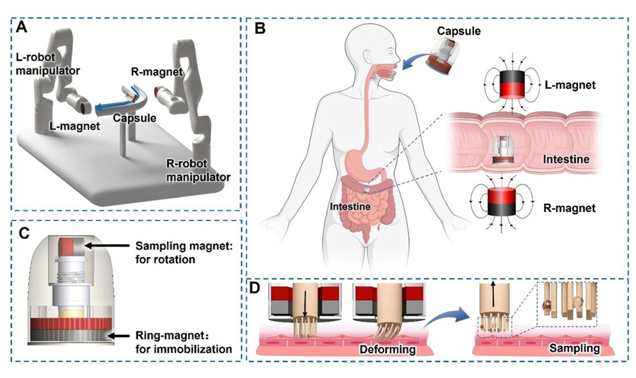
- Magnetically Guided Capsule Enables Targeted Small Intestine Microbiome Sampling
- Injectable Foam Dressing Conforms to Deep Traumatic Wounds and Inhibits Bacteria
- First Surgery Using Commercial Stem Cell-Derived Heart Muscle Sheets Performed in Japan
- New Software Tools Support Precision Mapping and Guidance in Cardiac Ablation
- Tile-Based Radiation Therapy Reduces Recurrence Risk After Brain Metastasis Surgery
- EHR Model Flags High-Risk Periods in Patients with Metastatic Breast Cancer
- Virtual Reality Helps Claustrophobic Patients Complete Cardiac PET/CT Scans
- Weighted Blankets Reduce Anxiety in Hospitalized Adults
- Multicenter Study Quantifies Delays in Care for Emergency Department Boarders
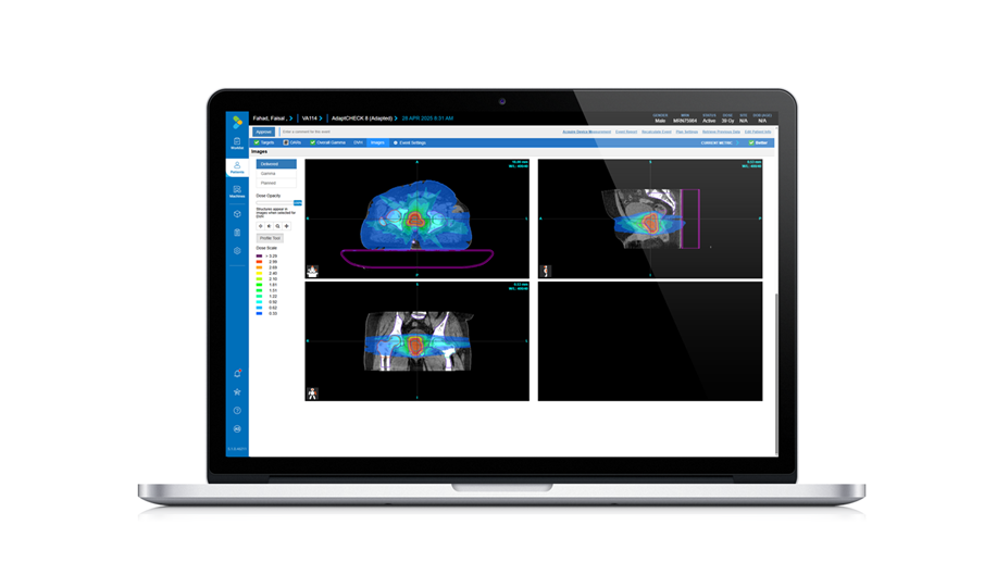
- Real-Time Radiation Monitor Supports Safer Fluoroscopy-Guided Procedures
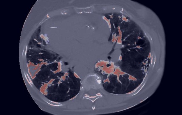
- AI Model Uses Pretreatment CT Scans to Predict Immunotherapy-Related Pneumonitis Risk
- MRI-Based Test Speeds Multiple Sclerosis Diagnosis and Reduces Lumbar Punctures
- AI Tool Improves Early Detection of Silicosis and Black Lung on X-Rays
- New MRI Approach Captures Rapidly Decaying Tissue Signals
- AI Tool Integrates MRI and Ultrasound to Detect Endometriosis
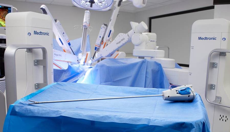
- Medtronic Receives FDA Clearance for Vessel-Sealing Instrument for Robotic Surgery
- Robotic Single-Port System Gains CE Mark for Transvaginal Gynecologic Procedures
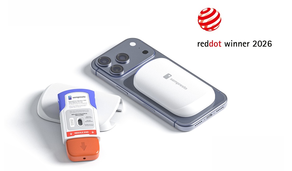
- Smartphone-Integrated Epinephrine Auto-Injector Concept Wins Red Dot Design Award
- First U.S. Commercial Cases Demonstrate New Approach to Transseptal Access
- FDA Approval Clears Path for BCI Integration with Personal Computing Devices
- Healthcare Database Helps Close Long COVID Surveillance Gap
- New Tablet Platform Extends Stroke Neurorehabilitation Beyond the Clinic
- Smartphone App Improves Blood Pressure Control in Hypertension
- Registry-Driven Digital Outreach Boosts Use of Heart Failure Medicines
- Nurse-Coordinated Care Model Improves Heart Failure Outcomes
- Autonomous AI Helps Streamline Urgent Skin Cancer Referrals
- Virtual Ultrasound Images Support Development of Cardiovascular Imaging Tools
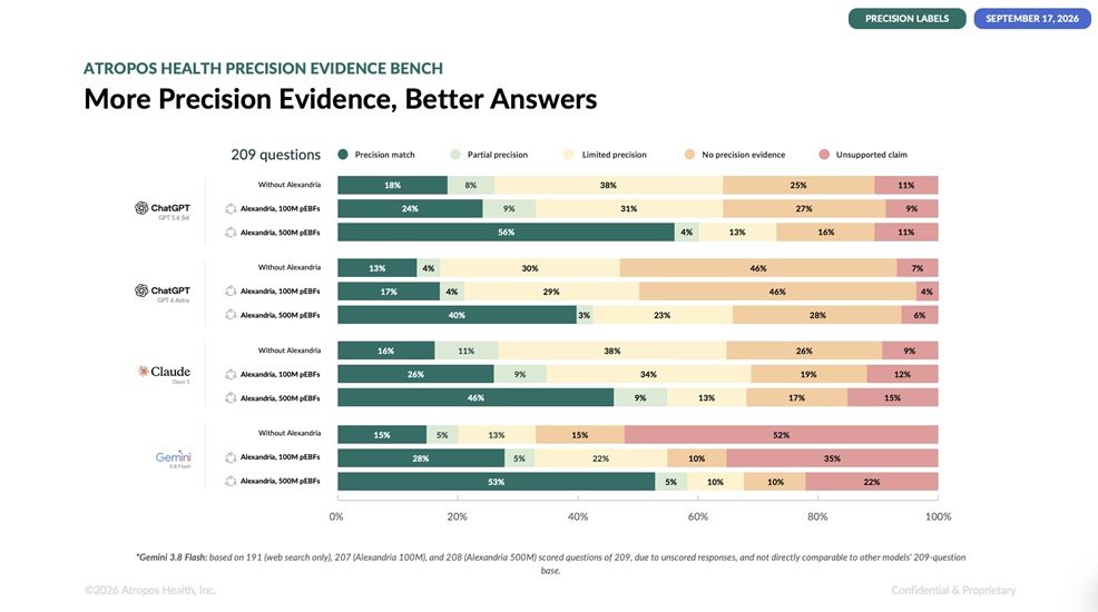
- New Benchmark Highlights Evidence Gaps in AI Responses to Clinical Questions
- Explainable AI Tool Improves Brain Disorder Screening from 4D fMRI
- AI Improves Non-Contrast CT Interpretation for Time-Sensitive Stroke Assessment
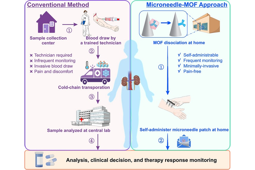
- Microneedle Patch Enables At-Home Monitoring of Acute Kidney Injury
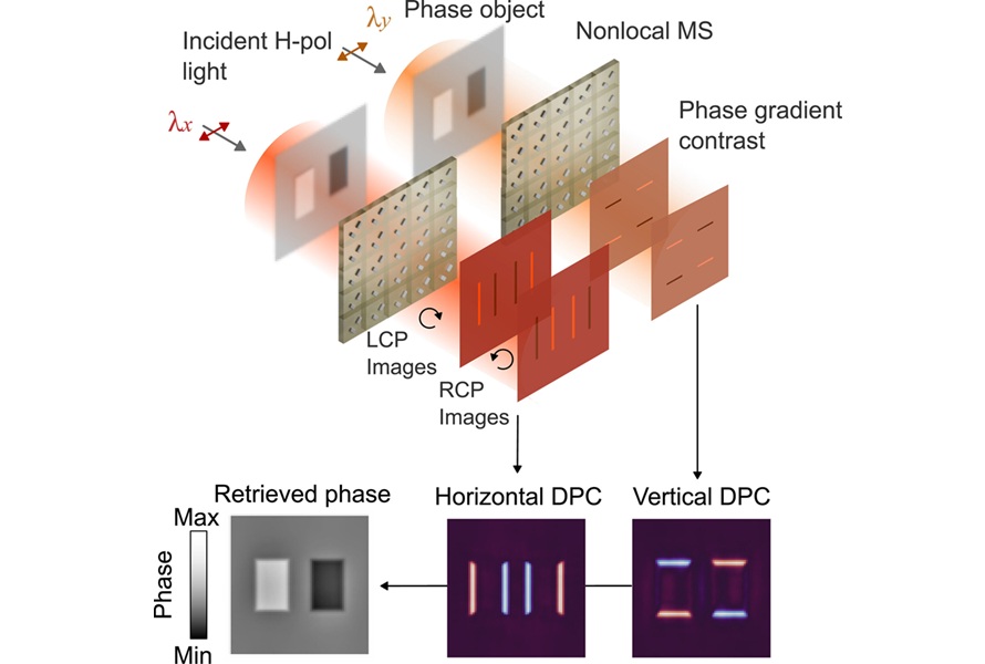
- Ultrathin Metasurface Could Bring Quantitative Phase Imaging to Portable Devices
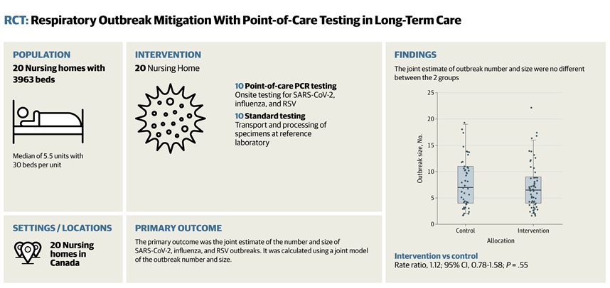
- Rapid Onsite Testing Reduces Emergency Department Transfers in Long-Term Care
- New Brain Ultrasound Platform Enables Bedside Postoperative Imaging
- Handheld Ultrasound Expands Point-of-Care Imaging Access in Brazil
 Expo
- Battery-Free Wearable Sensor Enables Activity Recognition for Health Monitoring
- New ECG Foundation Model Enables Broad Cardiac Diagnosis and Risk Prediction
- Nasal Spray Provides Rapid Pain Relief for Children in Emergency Care
- Noninvasive AI Tool Maps Atrial Cardiomyopathy to Support Ablation Planning
- Bioelectronic Network Enables Devices to Communicate Through Body Tissue and Coordinate Therapy
- Magnetically Guided Capsule Enables Targeted Small Intestine Microbiome Sampling
- Injectable Foam Dressing Conforms to Deep Traumatic Wounds and Inhibits Bacteria
- First Surgery Using Commercial Stem Cell-Derived Heart Muscle Sheets Performed in Japan
- New Software Tools Support Precision Mapping and Guidance in Cardiac Ablation
- Tile-Based Radiation Therapy Reduces Recurrence Risk After Brain Metastasis Surgery
- EHR Model Flags High-Risk Periods in Patients with Metastatic Breast Cancer
- Virtual Reality Helps Claustrophobic Patients Complete Cardiac PET/CT Scans
- Weighted Blankets Reduce Anxiety in Hospitalized Adults
- Multicenter Study Quantifies Delays in Care for Emergency Department Boarders
- Real-Time Radiation Monitor Supports Safer Fluoroscopy-Guided Procedures
- AI Model Uses Pretreatment CT Scans to Predict Immunotherapy-Related Pneumonitis Risk
- MRI-Based Test Speeds Multiple Sclerosis Diagnosis and Reduces Lumbar Punctures
- AI Tool Improves Early Detection of Silicosis and Black Lung on X-Rays
- New MRI Approach Captures Rapidly Decaying Tissue Signals
- AI Tool Integrates MRI and Ultrasound to Detect Endometriosis
- Medtronic Receives FDA Clearance for Vessel-Sealing Instrument for Robotic Surgery
- Robotic Single-Port System Gains CE Mark for Transvaginal Gynecologic Procedures
- Smartphone-Integrated Epinephrine Auto-Injector Concept Wins Red Dot Design Award
- First U.S. Commercial Cases Demonstrate New Approach to Transseptal Access
- FDA Approval Clears Path for BCI Integration with Personal Computing Devices
- Healthcare Database Helps Close Long COVID Surveillance Gap
- New Tablet Platform Extends Stroke Neurorehabilitation Beyond the Clinic
- Smartphone App Improves Blood Pressure Control in Hypertension
- Registry-Driven Digital Outreach Boosts Use of Heart Failure Medicines
- Nurse-Coordinated Care Model Improves Heart Failure Outcomes
- Autonomous AI Helps Streamline Urgent Skin Cancer Referrals
- Virtual Ultrasound Images Support Development of Cardiovascular Imaging Tools
- New Benchmark Highlights Evidence Gaps in AI Responses to Clinical Questions
- Explainable AI Tool Improves Brain Disorder Screening from 4D fMRI
- AI Improves Non-Contrast CT Interpretation for Time-Sensitive Stroke Assessment
- Microneedle Patch Enables At-Home Monitoring of Acute Kidney Injury
- Ultrathin Metasurface Could Bring Quantitative Phase Imaging to Portable Devices
- Rapid Onsite Testing Reduces Emergency Department Transfers in Long-Term Care
- New Brain Ultrasound Platform Enables Bedside Postoperative Imaging
- Handheld Ultrasound Expands Point-of-Care Imaging Access in Brazil































 Client Login