
Expo
view channel
view channel
view channel
view channel
view channel
view channel
view channel
Medical Imaging
AICritical Care
Patient CareHealth ITPoint of CareBusiness
Events
Webinars

- Invisible Skin Sensors Could Transform Everyday Biosignal Monitoring
- Patient-Specific Artery-on-a-Chip Refines Stroke Risk Stratification
- Existing Cardiovascular Risk Calculators Predict Several Common Cancers
- New Neurovascular Guidewire Enhances Access and Control in Stroke Thrombectomy
- AI Tool Automates EEG Analysis and Flags Clinically Relevant Abnormalities
- Navigation-Compatible Instruments Improve Precision in Lumbar Facet Fusion
- Reusable Pulse Oximetry Sensor Improves Accuracy Across Skin Tones
- Implantable Neural Interface Undergoes First Real-World Surgical Validation
- Robot-Guided Surgery Treats Multiple Deep Brain Abscesses in Single Session
- AI Surgical Platform Enables Real-Time Intraoperative Decision Support
- Wearable Sleep Data Predict Adherence to Pulmonary Rehabilitation
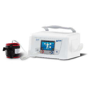
- Revolutionary Automatic IV-Line Flushing Device to Enhance Infusion Care
- VR Training Tool Combats Contamination of Portable Medical Equipment
- Portable Biosensor Platform to Reduce Hospital-Acquired Infections
- First-Of-Its-Kind Portable Germicidal Light Technology Disinfects High-Touch Clinical Surfaces in Seconds
- Danaher Completes Acquisition of Masimo to Expand Patient Monitoring Capabilities
- Endologix Adds FDA-Cleared Peripheral Thrombectomy System
- Artivion Adds FDA-Approved NEXUS System to Aortic Arch Portfolio
- Olympus Partnership Aims to Expand Access to Robot-Assisted Endoscopic Therapy
- Johnson & Johnson Launches AI-Driven Cardiac Mapping System
- Weekly Remote Symptom Monitoring Improves Symptom Control in Advanced Cancer
- Digital Heart Model Supports Targeted Ablation in Atrial Fibrillation
- AI Framework Helps Clinicians Create Trustworthy Risk Prediction Tools
- AI Tool Screens for Primary Aldosteronism Using Routine EHR Data
- AI-Enabled ECG Software Predicts One-Year Atrial Fibrillation Risk
- AI Tool Predicts Chronic Kidney Disease Risk in Diabetes
- AI Trends Report Guides Responsible, Effective Healthcare Deployment
- Privacy-Preserving AI Protects Sensitive Information in ECG Data
- New AI ECG Tool Detects Early Heart Disease
- AI Platform Supports Noninvasive Remote Hemodynamic Monitoring in Heart Failure
- Handheld Ultrasound Expands Point-of-Care Imaging Access in Brazil
- AI Dermatology Platform Targets Early Detection of Non-Melanoma Skin Cancer
- Handheld AI Device for Point-of-Care Skin Lesion Assessment Receives CE Mark
- Portable Immunoassay System Advances Toward Point-of-Care Biomarker Testing
- Portable MRI System Accelerates Emergency Brain Imaging and Triage
- Invisible Skin Sensors Could Transform Everyday Biosignal Monitoring
- Patient-Specific Artery-on-a-Chip Refines Stroke Risk Stratification
- Existing Cardiovascular Risk Calculators Predict Several Common Cancers
- New Neurovascular Guidewire Enhances Access and Control in Stroke Thrombectomy
- AI Tool Automates EEG Analysis and Flags Clinically Relevant Abnormalities
- Navigation-Compatible Instruments Improve Precision in Lumbar Facet Fusion
- Reusable Pulse Oximetry Sensor Improves Accuracy Across Skin Tones
- Implantable Neural Interface Undergoes First Real-World Surgical Validation
- Robot-Guided Surgery Treats Multiple Deep Brain Abscesses in Single Session
- AI Surgical Platform Enables Real-Time Intraoperative Decision Support
- Wearable Sleep Data Predict Adherence to Pulmonary Rehabilitation
- Revolutionary Automatic IV-Line Flushing Device to Enhance Infusion Care
- VR Training Tool Combats Contamination of Portable Medical Equipment
- Portable Biosensor Platform to Reduce Hospital-Acquired Infections
- First-Of-Its-Kind Portable Germicidal Light Technology Disinfects High-Touch Clinical Surfaces in Seconds
- Danaher Completes Acquisition of Masimo to Expand Patient Monitoring Capabilities
- Endologix Adds FDA-Cleared Peripheral Thrombectomy System
- Artivion Adds FDA-Approved NEXUS System to Aortic Arch Portfolio
- Olympus Partnership Aims to Expand Access to Robot-Assisted Endoscopic Therapy
- Johnson & Johnson Launches AI-Driven Cardiac Mapping System
- Weekly Remote Symptom Monitoring Improves Symptom Control in Advanced Cancer
- Digital Heart Model Supports Targeted Ablation in Atrial Fibrillation
- AI Framework Helps Clinicians Create Trustworthy Risk Prediction Tools
- AI Tool Screens for Primary Aldosteronism Using Routine EHR Data
- AI-Enabled ECG Software Predicts One-Year Atrial Fibrillation Risk
- AI Tool Predicts Chronic Kidney Disease Risk in Diabetes
- AI Trends Report Guides Responsible, Effective Healthcare Deployment
- Privacy-Preserving AI Protects Sensitive Information in ECG Data
- New AI ECG Tool Detects Early Heart Disease
- AI Platform Supports Noninvasive Remote Hemodynamic Monitoring in Heart Failure
- Handheld Ultrasound Expands Point-of-Care Imaging Access in Brazil
- AI Dermatology Platform Targets Early Detection of Non-Melanoma Skin Cancer
- Handheld AI Device for Point-of-Care Skin Lesion Assessment Receives CE Mark
- Portable Immunoassay System Advances Toward Point-of-Care Biomarker Testing
- Portable MRI System Accelerates Emergency Brain Imaging and Triage


")
")
")
 Model development using VAE with adversarial training to remove demographic attributes from ECGs. (B) Performance analysis for downstream tasks. (C) Applications of the privacy-preserving ECG embeddings in clinical outcome prediction and secure data sharing. (Fairuz Shadmani Shishir et al., Scientific Reports (2026). DOI: 10.1038/s41598-026-47665-6)")
")
 and the invisible electrode (right side) attached to the face without and with annotation (Photo courtesy of the Institute of Industrial Science, The University of Tokyo)")
. DOI: 10.1016/j.celbio.2026.100541)")

")
")
")
")


. DOI: 10.1113/jp290765)")
. (Photo courtesy of Longeviti Neuro Solutions)")
")
")
")
")


