





Expo
view channel
view channel
view channel
view channel
view channel
view channel
view channel
view channel
AICritical CareSurgical TechniquesPatient CareMedical ImagingHealth ITPoint of CareBusiness
Events
Webinars

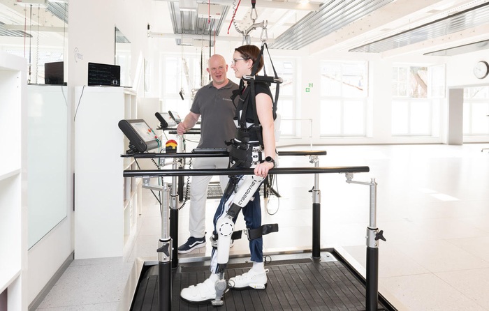
- Wearable Robotic Gait Training Linked to Improved Walking Distance in ALS
- Consumer Smartwatch May Expand Access to Obstructive Sleep Apnea Screening
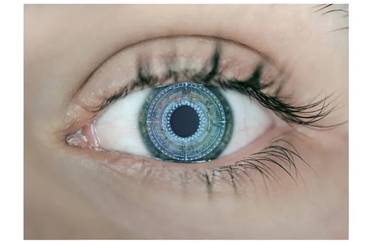
- Smart Tear Sensors May Help Detect Shifts in Eye Health Over Time
- Wearable Smart Ring Receives FDA Clearance for Noninvasive Blood Oxygen Monitoring
- Wristwatch Sensor Tracks Sodium and Potassium in Sweat in Real Time
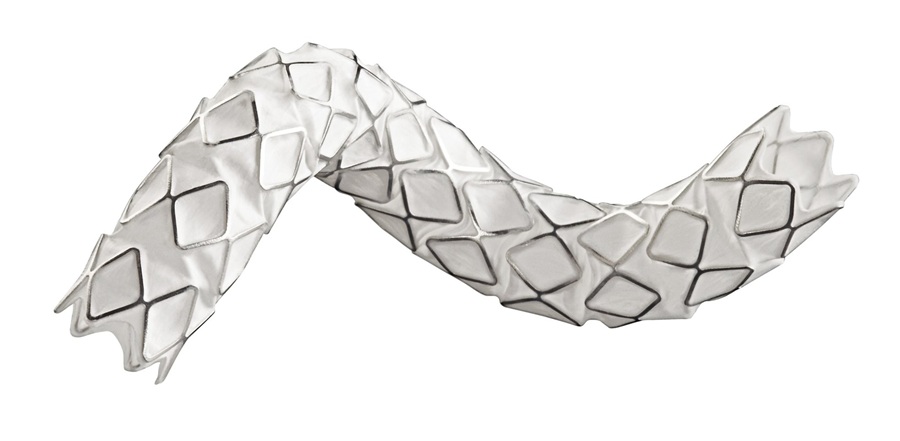
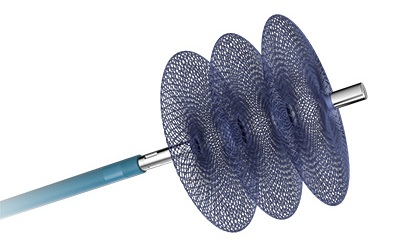
- FDA Approval Expands Bridging Stent Use in Complex Aortic Aneurysm Repair
- New Beam Measurement Approach Could Support More Precise Ion Therapy
- Vascular Occlusion System Gains Expanded FDA Indication for Venous Embolization
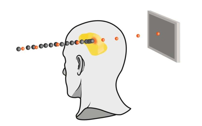
- Tiny Robots Could Speed Targeted Drug Delivery to Heart Infections
- Contact Radiotherapy Offers New Organ-Preserving Approach for Rectal Cancer
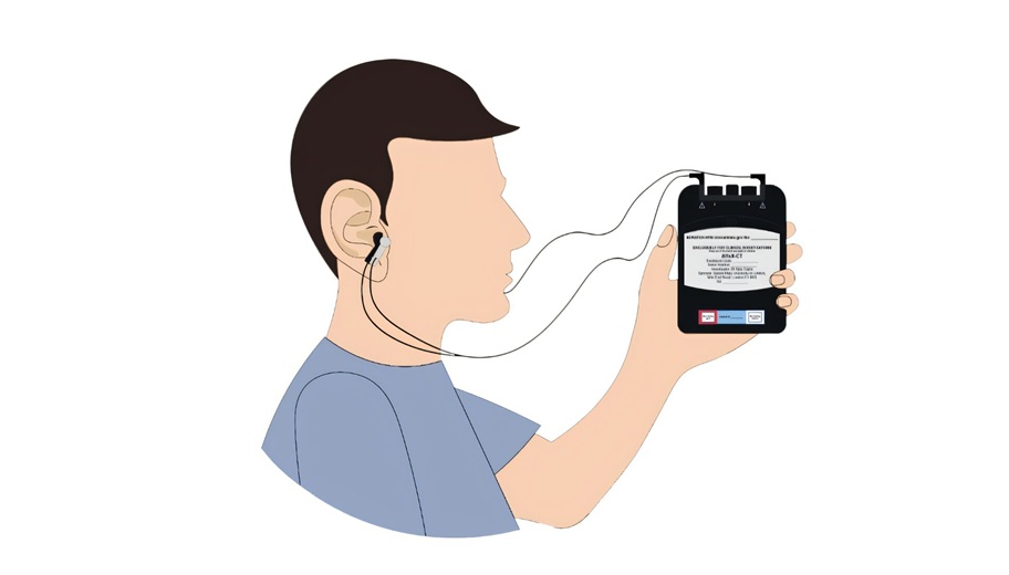
- At-Home Neuromodulation Shows Promise in Selected Patients With Hypertension
- EHR Model Flags High-Risk Periods in Patients with Metastatic Breast Cancer
- Virtual Reality Helps Claustrophobic Patients Complete Cardiac PET/CT Scans
- Weighted Blankets Reduce Anxiety in Hospitalized Adults
- Multicenter Study Quantifies Delays in Care for Emergency Department Boarders
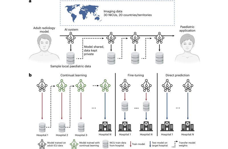
- Privacy-Preserving Learning Model Adapts Adult Radiology AI for Neonatal Care
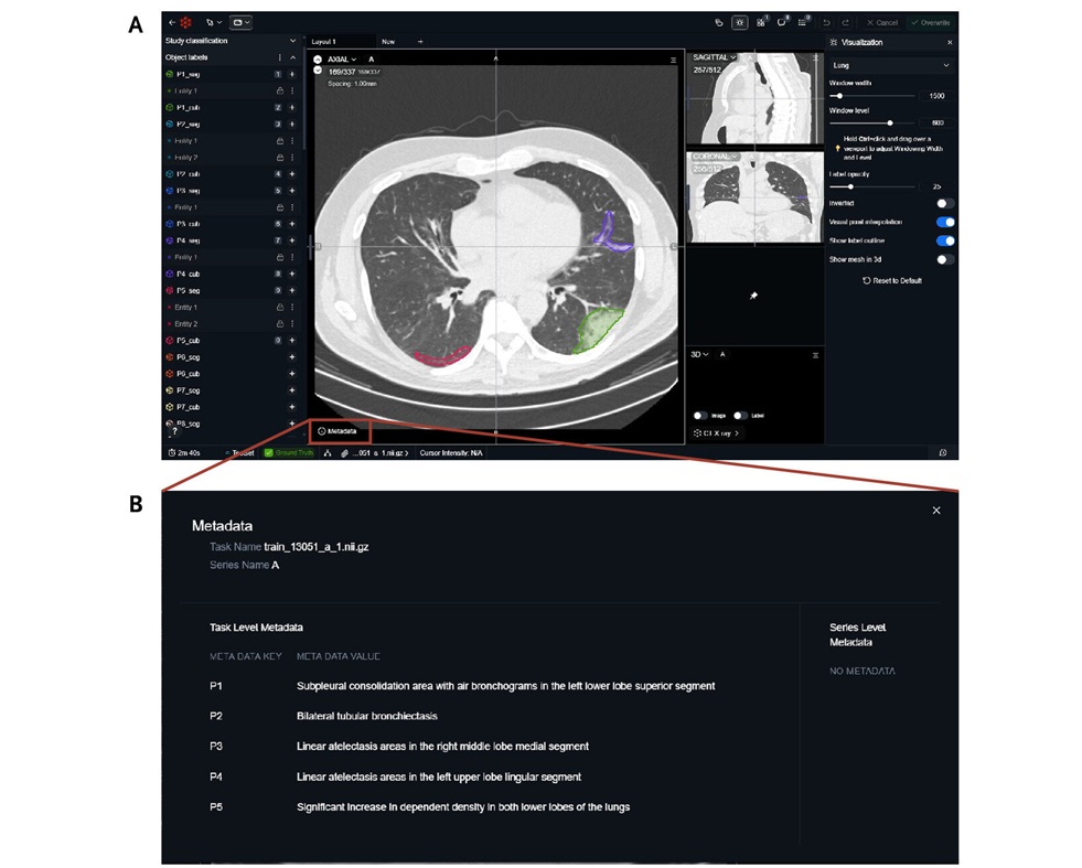
- Public Chest CT Dataset Supports Explainable AI in Diagnostic Radiology
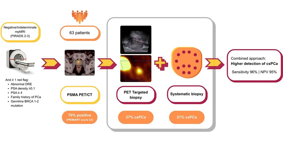
- PSMA PET/CT-Guided Biopsy Improves Detection of Significant Prostate Cancer After Nondiagnostic MRI
- AI Software Detects Osteoporosis from Routine CT Scans
- AI Model Uses Pretreatment CT Scans to Predict Immunotherapy-Related Pneumonitis Risk
- World Health Expo to Showcase Trillion-Dollar Growth Across Pharma, Biotech, Longevity and Wellness at 2027 Edition
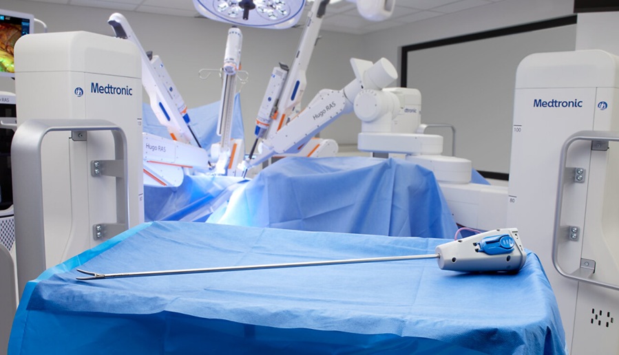
- Medtronic Receives FDA Clearance for Vessel-Sealing Instrument for Robotic Surgery
- Robotic Single-Port System Gains CE Mark for Transvaginal Gynecologic Procedures
- Smartphone-Integrated Epinephrine Auto-Injector Concept Wins Red Dot Design Award
- First U.S. Commercial Cases Demonstrate New Approach to Transseptal Access
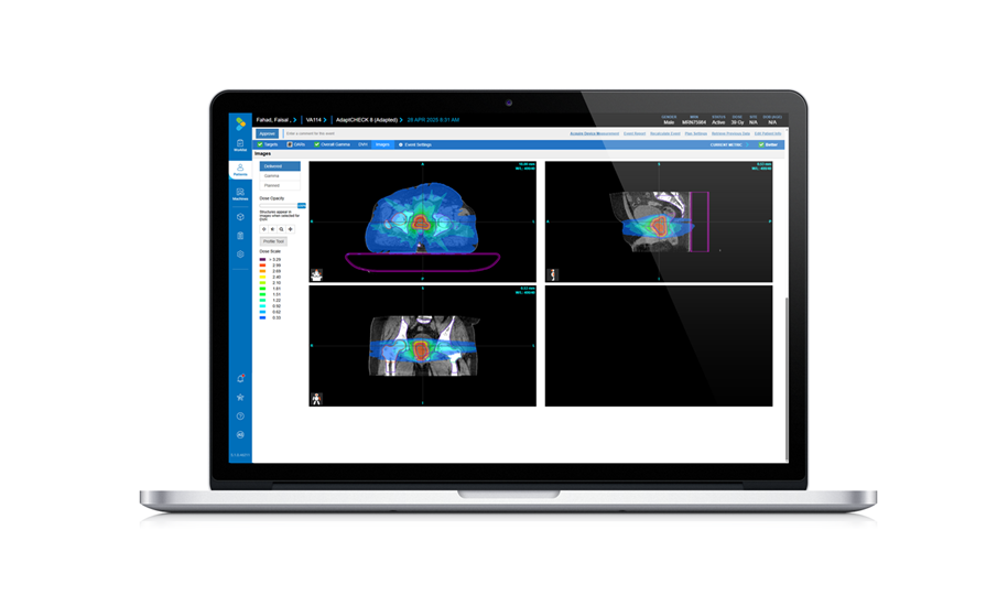
- Adaptive Radiation QA Software Gains Validation for NeoArc Treatments
- Healthcare Database Helps Close Long COVID Surveillance Gap
- New Tablet Platform Extends Stroke Neurorehabilitation Beyond the Clinic
- Smartphone App Improves Blood Pressure Control in Hypertension
- Registry-Driven Digital Outreach Boosts Use of Heart Failure Medicines
- AI Tool Predicts Long-Term Cardiovascular Risk from Sleep Study ECG
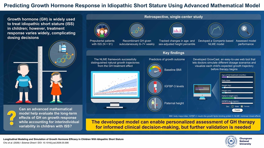
- Predictive Model Supports Growth Hormone Treatment Planning in Idiopathic Short Stature
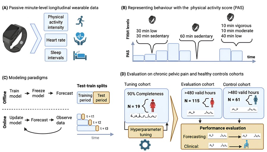
- AI Model Predicts Prolonged Sitting in Chronic Pelvic Pain
- Autonomous AI Helps Streamline Urgent Skin Cancer Referrals
- Virtual Ultrasound Images Support Development of Cardiovascular Imaging Tools
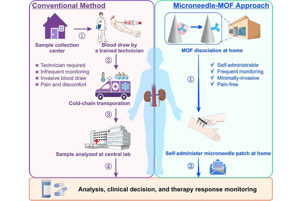
- Microneedle Patch Enables At-Home Monitoring of Acute Kidney Injury
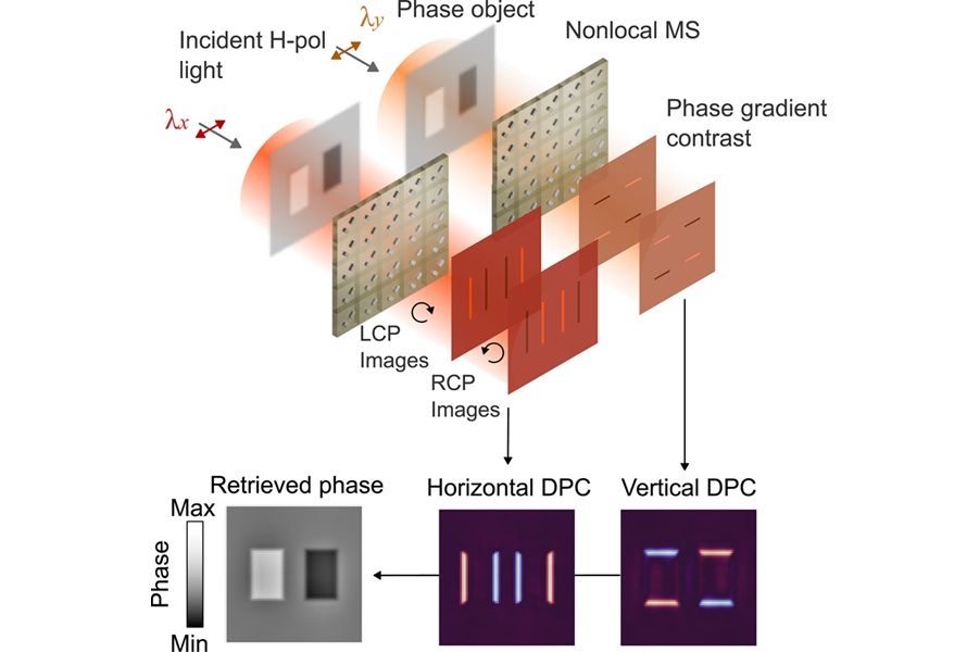
- Ultrathin Metasurface Could Bring Quantitative Phase Imaging to Portable Devices
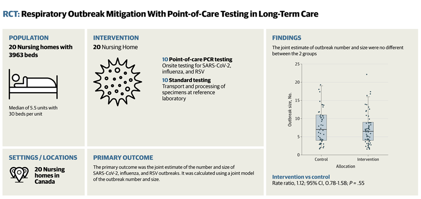
- Rapid Onsite Testing Reduces Emergency Department Transfers in Long-Term Care
- New Brain Ultrasound Platform Enables Bedside Postoperative Imaging
- Handheld Ultrasound Expands Point-of-Care Imaging Access in Brazil
 Expo
Expo
- Wearable Robotic Gait Training Linked to Improved Walking Distance in ALS
- Consumer Smartwatch May Expand Access to Obstructive Sleep Apnea Screening
- Smart Tear Sensors May Help Detect Shifts in Eye Health Over Time
- Wearable Smart Ring Receives FDA Clearance for Noninvasive Blood Oxygen Monitoring
- Wristwatch Sensor Tracks Sodium and Potassium in Sweat in Real Time
- FDA Approval Expands Bridging Stent Use in Complex Aortic Aneurysm Repair
- New Beam Measurement Approach Could Support More Precise Ion Therapy
- Vascular Occlusion System Gains Expanded FDA Indication for Venous Embolization
- Tiny Robots Could Speed Targeted Drug Delivery to Heart Infections
- Contact Radiotherapy Offers New Organ-Preserving Approach for Rectal Cancer
- At-Home Neuromodulation Shows Promise in Selected Patients With Hypertension
- EHR Model Flags High-Risk Periods in Patients with Metastatic Breast Cancer
- Virtual Reality Helps Claustrophobic Patients Complete Cardiac PET/CT Scans
- Weighted Blankets Reduce Anxiety in Hospitalized Adults
- Multicenter Study Quantifies Delays in Care for Emergency Department Boarders
- Privacy-Preserving Learning Model Adapts Adult Radiology AI for Neonatal Care
- Public Chest CT Dataset Supports Explainable AI in Diagnostic Radiology
- PSMA PET/CT-Guided Biopsy Improves Detection of Significant Prostate Cancer After Nondiagnostic MRI
- AI Software Detects Osteoporosis from Routine CT Scans
- AI Model Uses Pretreatment CT Scans to Predict Immunotherapy-Related Pneumonitis Risk
- World Health Expo to Showcase Trillion-Dollar Growth Across Pharma, Biotech, Longevity and Wellness at 2027 Edition
- Medtronic Receives FDA Clearance for Vessel-Sealing Instrument for Robotic Surgery
- Robotic Single-Port System Gains CE Mark for Transvaginal Gynecologic Procedures
- Smartphone-Integrated Epinephrine Auto-Injector Concept Wins Red Dot Design Award
- First U.S. Commercial Cases Demonstrate New Approach to Transseptal Access
- Adaptive Radiation QA Software Gains Validation for NeoArc Treatments
- Healthcare Database Helps Close Long COVID Surveillance Gap
- New Tablet Platform Extends Stroke Neurorehabilitation Beyond the Clinic
- Smartphone App Improves Blood Pressure Control in Hypertension
- Registry-Driven Digital Outreach Boosts Use of Heart Failure Medicines
- AI Tool Predicts Long-Term Cardiovascular Risk from Sleep Study ECG
- Predictive Model Supports Growth Hormone Treatment Planning in Idiopathic Short Stature
- AI Model Predicts Prolonged Sitting in Chronic Pelvic Pain
- Autonomous AI Helps Streamline Urgent Skin Cancer Referrals
- Virtual Ultrasound Images Support Development of Cardiovascular Imaging Tools
- Microneedle Patch Enables At-Home Monitoring of Acute Kidney Injury
- Ultrathin Metasurface Could Bring Quantitative Phase Imaging to Portable Devices
- Rapid Onsite Testing Reduces Emergency Department Transfers in Long-Term Care
- New Brain Ultrasound Platform Enables Bedside Postoperative Imaging
- Handheld Ultrasound Expands Point-of-Care Imaging Access in Brazil


































 Client Login