





Expo
view channel
view channel
view channel
view channel
view channel
view channel
view channel
Medical Imaging
AI
Surgical TechniquesPatient CareHealth ITPoint of CareBusiness
Events
Webinars

- New Neurovascular Guidewire Enhances Access and Control in Stroke Thrombectomy
- AI Tool Automates EEG Analysis and Flags Clinically Relevant Abnormalities
- Low-Cost Antiseptic Regimen Reduces Bacteria Linked to Newborn Infections
- New Risk Models Improve Sudden Cardiac Arrest Prediction
- Collaborative ICU Admission Process Cuts Time to Critical Care Involvement by Half
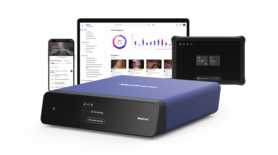
- AI Surgical Platform Enables Real-Time Intraoperative Decision Support
- AI-Guided Arterial Mapping Improves Brain Tumor Drug Delivery
- Implanted Nerve Stimulation May Improve Health Outcomes in Sleep Apnea
- AI-Designed Microneedle Patch Accelerates Diabetic Wound Healing
- Focused Ultrasound System Gains CE Mark for Liver Tumor Treatment
- Wearable Sleep Data Predict Adherence to Pulmonary Rehabilitation
- Revolutionary Automatic IV-Line Flushing Device to Enhance Infusion Care
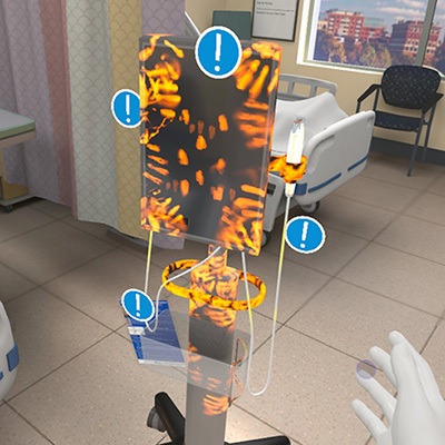
- VR Training Tool Combats Contamination of Portable Medical Equipment
- Portable Biosensor Platform to Reduce Hospital-Acquired Infections
- First-Of-Its-Kind Portable Germicidal Light Technology Disinfects High-Touch Clinical Surfaces in Seconds
- Danaher Completes Acquisition of Masimo to Expand Patient Monitoring Capabilities
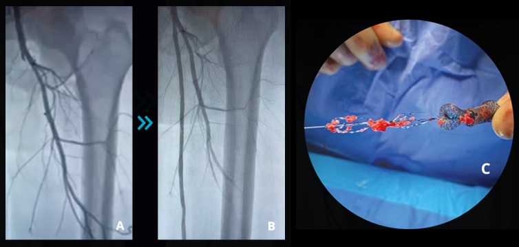
- Endologix Adds FDA-Cleared Peripheral Thrombectomy System
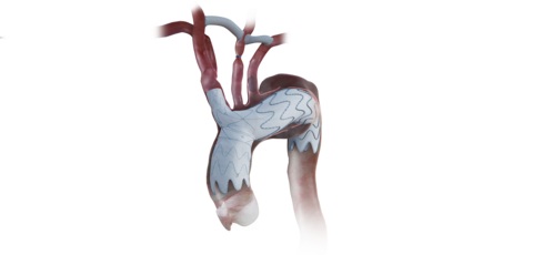
- Artivion Adds FDA-Approved NEXUS System to Aortic Arch Portfolio
- Olympus Partnership Aims to Expand Access to Robot-Assisted Endoscopic Therapy
- Johnson & Johnson Launches AI-Driven Cardiac Mapping System
- Weekly Remote Symptom Monitoring Improves Symptom Control in Advanced Cancer
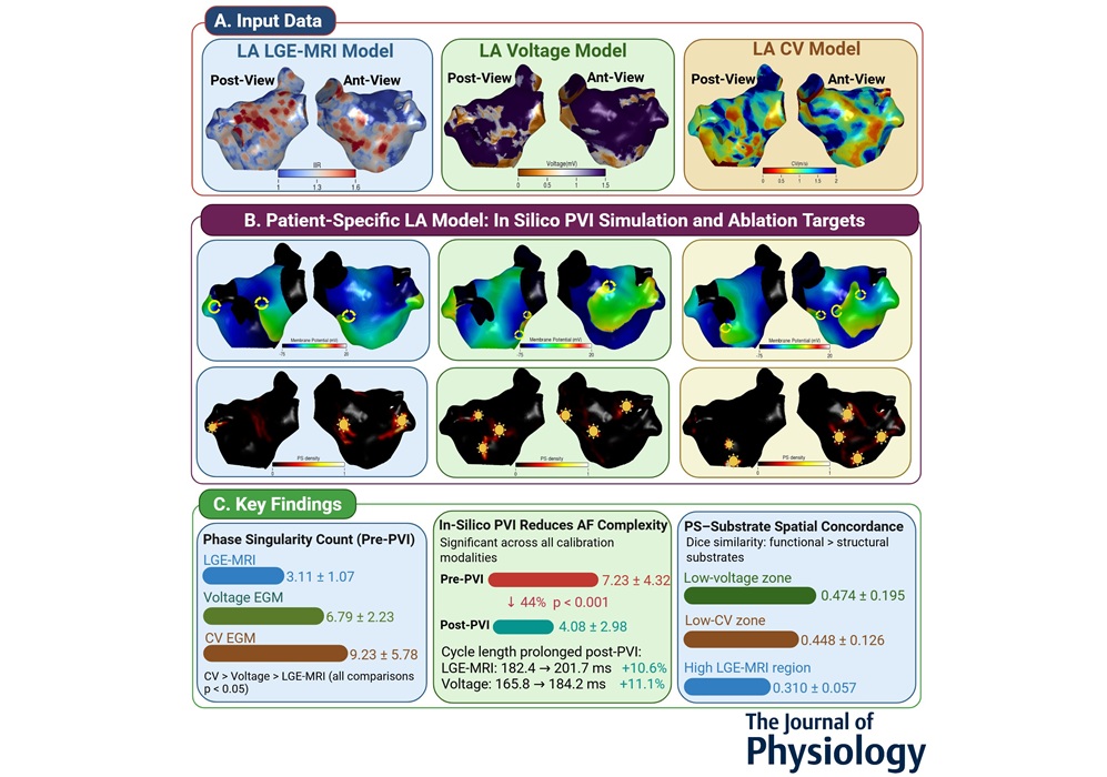
- Digital Heart Model Supports Targeted Ablation in Atrial Fibrillation
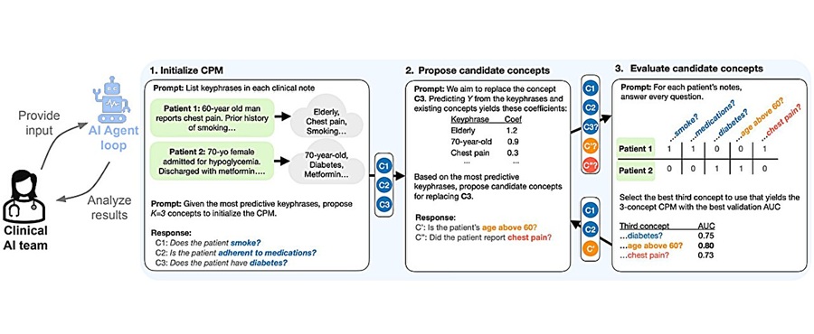
- AI Framework Helps Clinicians Create Trustworthy Risk Prediction Tools
- AI Tool Screens for Primary Aldosteronism Using Routine EHR Data
- AI-Enabled ECG Software Predicts One-Year Atrial Fibrillation Risk
- AI Tool Predicts Chronic Kidney Disease Risk in Diabetes
- AI Trends Report Guides Responsible, Effective Healthcare Deployment
- Privacy-Preserving AI Protects Sensitive Information in ECG Data
- New AI ECG Tool Detects Early Heart Disease
- AI Platform Supports Noninvasive Remote Hemodynamic Monitoring in Heart Failure
- Handheld Ultrasound Expands Point-of-Care Imaging Access in Brazil
- AI Dermatology Platform Targets Early Detection of Non-Melanoma Skin Cancer
- Handheld AI Device for Point-of-Care Skin Lesion Assessment Receives CE Mark
- Portable Immunoassay System Advances Toward Point-of-Care Biomarker Testing
- Portable MRI System Accelerates Emergency Brain Imaging and Triage
 Expo
Expo
- New Neurovascular Guidewire Enhances Access and Control in Stroke Thrombectomy
- AI Tool Automates EEG Analysis and Flags Clinically Relevant Abnormalities
- Low-Cost Antiseptic Regimen Reduces Bacteria Linked to Newborn Infections
- New Risk Models Improve Sudden Cardiac Arrest Prediction
- Collaborative ICU Admission Process Cuts Time to Critical Care Involvement by Half
- AI Surgical Platform Enables Real-Time Intraoperative Decision Support
- AI-Guided Arterial Mapping Improves Brain Tumor Drug Delivery
- Implanted Nerve Stimulation May Improve Health Outcomes in Sleep Apnea
- AI-Designed Microneedle Patch Accelerates Diabetic Wound Healing
- Focused Ultrasound System Gains CE Mark for Liver Tumor Treatment
- Wearable Sleep Data Predict Adherence to Pulmonary Rehabilitation
- Revolutionary Automatic IV-Line Flushing Device to Enhance Infusion Care
- VR Training Tool Combats Contamination of Portable Medical Equipment
- Portable Biosensor Platform to Reduce Hospital-Acquired Infections
- First-Of-Its-Kind Portable Germicidal Light Technology Disinfects High-Touch Clinical Surfaces in Seconds
- Danaher Completes Acquisition of Masimo to Expand Patient Monitoring Capabilities
- Endologix Adds FDA-Cleared Peripheral Thrombectomy System
- Artivion Adds FDA-Approved NEXUS System to Aortic Arch Portfolio
- Olympus Partnership Aims to Expand Access to Robot-Assisted Endoscopic Therapy
- Johnson & Johnson Launches AI-Driven Cardiac Mapping System
- Weekly Remote Symptom Monitoring Improves Symptom Control in Advanced Cancer
- Digital Heart Model Supports Targeted Ablation in Atrial Fibrillation
- AI Framework Helps Clinicians Create Trustworthy Risk Prediction Tools
- AI Tool Screens for Primary Aldosteronism Using Routine EHR Data
- AI-Enabled ECG Software Predicts One-Year Atrial Fibrillation Risk
- AI Tool Predicts Chronic Kidney Disease Risk in Diabetes
- AI Trends Report Guides Responsible, Effective Healthcare Deployment
- Privacy-Preserving AI Protects Sensitive Information in ECG Data
- New AI ECG Tool Detects Early Heart Disease
- AI Platform Supports Noninvasive Remote Hemodynamic Monitoring in Heart Failure
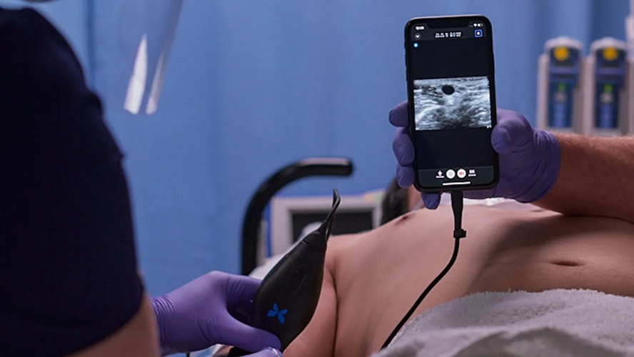
- Handheld Ultrasound Expands Point-of-Care Imaging Access in Brazil
- AI Dermatology Platform Targets Early Detection of Non-Melanoma Skin Cancer
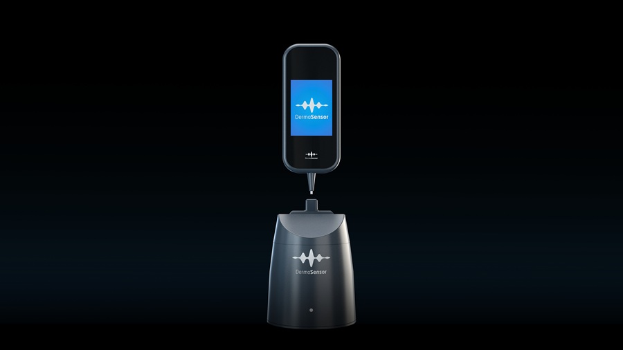
- Handheld AI Device for Point-of-Care Skin Lesion Assessment Receives CE Mark
- Portable Immunoassay System Advances Toward Point-of-Care Biomarker Testing
- Portable MRI System Accelerates Emergency Brain Imaging and Triage





























 Client Login