We use cookies to understand how you use our site and to improve your experience. This includes personalizing content and advertising. To learn more, click here. By continuing to use our site, you accept our use of cookies. Cookie Policy.

Features Partner Sites Information LinkXpress hp
Sign In
Advertise with Us
ARAB HEALTH - INFORMA

Download Mobile App


Parametric Modeling Helps Determine Prosthetic Valve Size

By HospiMedica International staff writers
Posted on 02 Jan 2019
Print article
Image: Physical 3D printed models of patient aortic heart valves (Photo courtesy of Wyss Institute).
A new study describes how three-dimensional (3D) printing can evaluate how different valve sizes will interact with each patient's unique anatomy, before the procedure is actually performed.

Developed by researchers at the Max Planck Institute of Colloids and Interfaces (MPIKG; Potsdam, Germany), the Wyss Institute for Biologically Inspired Engineering (Boston, MA, USA), Massachusetts General Hospital (MGH; Boston, USA), and other institutions, the software program uses parametric modeling to generate virtual 3D models of the leaflets, using seven calcification coordinates visible on computerized tomography (CT) scans. The resulting model, which incorporates both leaflets and their associated calcified deposits, is then 3D printed.

The 3D-printed multi-material valve model incorporates flexible leaflets and rigid calcified deposits that mimic the artificial valve deployment, as well as providing haptic feedback. A custom sizer that fits inside the 3D-printed valve model is also printed and wrapped with a thin layer of pressure-sensing film to map the contacts between the sizer and the 3D-printed valves and their associated calcified deposits. The sizer is gradually expanded until the correct fit is achieved. Subsequently, the researchers conducted a retrospective study of 30 patients who underwent transcatheter aortic valve replacement (TAVR).

3D printed adjustable sizers were then positioned in the aortic root models and sequentially opened to larger valve sizes, progressively flattening the calcified leaflets against the aortic wall. Optimal valve size and fit were determined by visual inspection and quantitative pressure mapping of interactions between the sizer and models. The researchers found that pressure testing provided a physical map of areas with an inadequate seal that corresponded to areas of paravalvular leak, as demonstrated by post-procedural transthoracic echocardiogram (TTE). The study was published on October 2, 2018, in the Journal of Cardiovascular Computed Tomography.

“If you buy a pair of shoes online without trying them on first, there's a good chance they're not going to fit properly. Sizing replacement TAVR valves poses a similar problem, in that doctors don't get the opportunity to evaluate how a specific valve size will fit with a patient's anatomy before surgery,” said corresponding author James Weaver, PhD, of the Wyss Institute. “Our integrative 3D printing and valve sizing system provides a customized report of every patient's unique aortic valve shape, removing a lot of the guesswork and helping each patient receive a more accurately sized valve.”

“Being able to identify intermediate- and low-risk patients whose heart valve anatomy gives them a higher probability of complications from TAVR is critical, and we've never had a non-invasive way to accurately determine that before,” said study co-author Beth Ripley, MD, PhD, of the University of Washington (Seattle, USA). “Those patients might be better served by surgery, as the risks of an imperfect TAVR result might outweigh its benefits. Additionally, being able to physically simulate the procedure might inform future iterations of valve designs and deployment approaches.”

The leaflet modeling software and the 3D printing protocol are freely available online for researchers or clinicians who wish to use them.

Related Links:
Max Planck Institute of Colloids and Interfaces
Wyss Institute for Biologically Inspired Engineering
Massachusetts General Hospital

Gold Member
12-Channel ECG
CM1200B
Gold Member
POC Blood Gas Analyzer
Stat Profile Prime Plus
New
Digital Radiographic System
OMNERA 300M
New
Diagnostic Ultrasound System
MS1700C

Print article

Channels

Critical Care

view channel
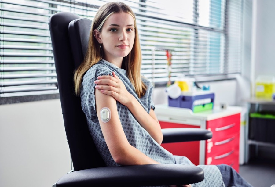
Image: Various sensors might be helpful at different ages (Photo courtesy of Brasier et al./Nature, 2024)

New Generation of Wearable Sensors to Perform Biochemical Analysis of Body Fluids

Wearable devices are already capable of monitoring vital body functions, such as pulse with a smartwatch or blood pressure with a smartphone app. While these sensors can provide reliable real-time data... Read more

Surgical Techniques

view channel
Image: Synthetic images generated by each diffusion model contrasted with the corresponding real textural images of four types of polyps (Photo courtesy of UT at Austin)

AI-Assisted Imaging to Assist Endoscopists in Colonoscopy Procedures

Colorectal cancer is a major health concern in the United States, with the likelihood of developing the disease being 1 in 25 for women and 1 in 23 for men. Polyps, which are precursors to cancer, can... Read more

Patient Care

view channel
Image: The portable biosensor platform uses printed electrochemical sensors for the rapid, selective detection of Staphylococcus aureus (Photo courtesy of AIMPLAS)

Portable Biosensor Platform to Reduce Hospital-Acquired Infections

Approximately 4 million patients in the European Union acquire healthcare-associated infections (HAIs) or nosocomial infections each year, with around 37,000 deaths directly resulting from these infections,... Read more

Point of Care

view channel
Image: The acoustic pipette uses sound waves to test for biomarkers in blood (Photo courtesy of Patrick Campbell/CU Boulder)

Handheld, Sound-Based Diagnostic System Delivers Bedside Blood Test Results in An Hour

Patients who go to a doctor for a blood test often have to contend with a needle and syringe, followed by a long wait—sometimes hours or even days—for lab results. Scientists have been working hard to... Read more
Copyright © 2000-2024 Globetech Media. All rights reserved.